

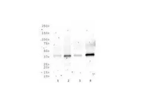
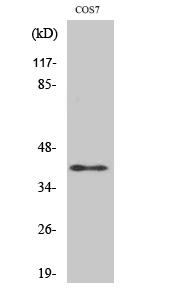
WB analysis of (1) HeLa, (2) HepG2, (3) COS-7, and (4) HEK293 cell lysate using GTX30137 Rad51C antibody [2H11/6].
![FACS (Intracellular staining) analysis of HeLa cells using GTX30137 Rad51C antibody [2H11/6]. Blue : Primary antibody Orange : isotype control Dilution : 1 μg/10? cells](https://www.genetex.com/upload/website/prouct_img/normal/GTX30137/GTX30137_95_FACS_w_23060722_355.webp "FACS (Intracellular staining) analysis of HeLa cells using GTX30137 Rad51C antibody [2H11/6]. Blue : Primary antibody Orange : isotype control Dilution : 1 μg/10? cells")
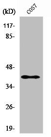
![WB analysis of HepG2 cell lysate using GTX30137 Rad51C antibody [2H11/6].](https://www.genetex.com/upload/website/prouct_img/normal/GTX30137/GTX30137_1342_WB_w_23060722_833.webp "WB analysis of HepG2 cell lysate using GTX30137 Rad51C antibody [2H11/6].")
WB analysis of (1) HeLa, (2) HepG2, (3) COS-7, and (4) HEK293 cell lysate using GTX30137 Rad51C antibody [2H11/6].
Rad51C antibody [2H11/6]
GTX30137
ApplicationsFlow Cytometry, ImmunoFluorescence, ImmunoPrecipitation, Mass Spectrometry, Western Blot, ImmunoCytoChemistry, ImmunoHistoChemistry, ImmunoHistoChemistry Paraffin
Product group Antibodies
ReactivityHuman, Monkey, Mouse, Primate, Yeast
TargetRAD51C
Overview
- SupplierGeneTex
- Product NameRad51C antibody [2H11/6]
- Delivery Days Customer9
- Application Supplier NoteWB: 1:1000. FCM: 1 microg / 106 cells. *Optimal dilutions/concentrations should be determined by the researcher.Not tested in other applications.
- ApplicationsFlow Cytometry, ImmunoFluorescence, ImmunoPrecipitation, Mass Spectrometry, Western Blot, ImmunoCytoChemistry, ImmunoHistoChemistry, ImmunoHistoChemistry Paraffin
- CertificationResearch Use Only
- ClonalityMonoclonal
- Clone ID2H11/6
- Concentration1 mg/ml
- ConjugateUnconjugated
- Gene ID5889
- Target nameRAD51C
- Target descriptionRAD51 paralog C
- Target synonymsBROVCA3, FANCO, R51H3, RAD51L2, DNA repair protein RAD51 homolog 3, RAD51-like protein 2, yeast RAD51 homolog 3
- HostMouse
- IsotypeIgG1
- Protein IDO43502
- Protein NameDNA repair protein RAD51 homolog 3
- Scientific DescriptionThis gene is a member of the RAD51 family of related genes, which encode strand-transfer proteins thought to be involved in recombinational repair of damaged DNA and in meiotic recombination. This gene product interacts with two other DNA repair proteins, encoded by RAD51B and XRCC3, but not with itself. The protein copurifies with XRCC3 protein in a complex, reflecting their endogenous association and suggesting a cooperative role during recombinational repair. This gene is one of four localized to a region of chromosome 17q23 where amplification occurs frequently in breast tumors. Overexpression of the four genes during amplification has been observed and suggests a possible role in tumor progression. Alternative splicing has been observed for this gene and two variants encoding different isoforms have been identified. [provided by RefSeq]
- ReactivityHuman, Monkey, Mouse, Primate, Yeast
- Storage Instruction2°C to 8°C
- UNSPSC41116161
Datasheet
Related products
Product group Antibodies
ApplicationsImmunoFluorescence, Western Blot, ImmunoHistoChemistry
ReactivityHuman, Mouse, Rat
- SizePrice
Product group Antibodies
Anti-RAD51C Antibody144-62348
ApplicationsImmunoFluorescence
ReactivityHuman
TargetRAD51C
- SizePrice
Product group Antibodies

Anti-Rad51C [2H11/6]Ab01304-1.1
ApplicationsImmunoFluorescence, ImmunoPrecipitation, Western Blot, ImmunoHistoChemistry
ReactivityHuman, Mouse, Primate, Yeast
TargetRAD51C
- SizePrice
Product group Antibodies

RAD51C AntibodyLS-C747585
ApplicationsImmunoFluorescence
ReactivityHuman
TargetRAD51C
- SizePrice

Product group Antibodies
Anti-RAD51C Antibody Picoband(r)A01837-1-CARRIER-FREE
ApplicationsFlow Cytometry, Western Blot, ELISA, ImmunoHistoChemistry
ReactivityHuman, Rat
TargetRAD51C
- SizePrice
Product group Antibodies
RAD51C AntibodyCSB-PA003917
ApplicationsImmunoFluorescence, Western Blot, ELISA, ImmunoHistoChemistry
ReactivityHuman, Monkey
TargetRAD51C
- SizePrice
Product group Antibodies

Goat anti-RAD51CEB05686
ApplicationsWestern Blot, ELISA
ReactivityHuman
TargetRAD51C
- SizePrice
Product group Antibodies
Rad51C antibody, N-termGTX89889
ApplicationsWestern Blot
ReactivityHuman
TargetRAD51C
- SizePrice

Product group Antibodies
Anti-RAD51C AntibodyHPA045198
ApplicationsImmunoCytoChemistry
ReactivityHuman
TargetRAD51C
- SizePrice
Product group Antibodies
Rad51C antibodyGTX34155
ApplicationsWestern Blot
ReactivityHuman, Monkey
TargetRAD51C
- SizePrice