

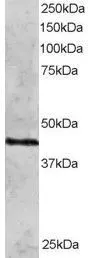
WB analysis of HeLa lysate using GTX89889 Rad51C antibody, N-term. Dilution : 2μg/ml Loading : 30μg protein in RIPA buffer
WB analysis of HeLa lysate using GTX89889 Rad51C antibody, N-term. Dilution : 2μg/ml Loading : 30μg protein in RIPA buffer
Rad51C antibody, N-term
GTX89889
ApplicationsWestern Blot
Product group Antibodies
ReactivityHuman
TargetRAD51C
Overview
- SupplierGeneTex
- Product NameRad51C antibody, N-term
- Delivery Days Customer9
- Application Supplier NoteWB: 1-3microg/ml. *Optimal dilutions/concentrations should be determined by the researcher.Not tested in other applications.
- ApplicationsWestern Blot
- CertificationResearch Use Only
- ClonalityPolyclonal
- Concentration0.50 mg/ml
- ConjugateUnconjugated
- Gene ID5889
- Target nameRAD51C
- Target descriptionRAD51 paralog C
- Target synonymsBROVCA3, FANCO, R51H3, RAD51L2, DNA repair protein RAD51 homolog 3, RAD51-like protein 2, yeast RAD51 homolog 3
- HostGoat
- IsotypeIgG
- Protein IDO43502
- Protein NameDNA repair protein RAD51 homolog 3
- Scientific DescriptionThis gene is a member of the RAD51 family. RAD51 family members are highly similar to bacterial RecA and Saccharomyces cerevisiae Rad51 and are known to be involved in the homologous recombination and repair of DNA. This protein can interact with other RAD51 paralogs and is reported to be important for Holliday junction resolution. Mutations in this gene are associated with Fanconi anemia-like syndrome. This gene is one of four localized to a region of chromosome 17q23 where amplification occurs frequently in breast tumors. Overexpression of the four genes during amplification has been observed and suggests a possible role in tumor progression. Alternative splicing results in multiple transcript variants. [provided by RefSeq, Jul 2013]
- ReactivityHuman
- Storage Instruction-20°C or -80°C,2°C to 8°C
- UNSPSC41116161
Datasheet
Related products
Product group Antibodies
ApplicationsImmunoFluorescence, Western Blot, ImmunoHistoChemistry
ReactivityHuman, Mouse, Rat
- SizePrice
Product group Antibodies
Anti-RAD51C Antibody144-62348
ApplicationsImmunoFluorescence
ReactivityHuman
TargetRAD51C
- SizePrice
Product group Antibodies

Anti-Rad51C [2H11/6]Ab01304-1.1
ApplicationsImmunoFluorescence, ImmunoPrecipitation, Western Blot, ImmunoHistoChemistry
ReactivityHuman, Mouse, Primate, Yeast
TargetRAD51C
- SizePrice
Product group Antibodies

RAD51C AntibodyLS-C747585
ApplicationsImmunoFluorescence
ReactivityHuman
TargetRAD51C
- SizePrice

Product group Antibodies
Anti-RAD51C Antibody Picoband(r)A01837-1-CARRIER-FREE
ApplicationsFlow Cytometry, Western Blot, ELISA, ImmunoHistoChemistry
ReactivityHuman, Rat
TargetRAD51C
- SizePrice
Product group Antibodies
RAD51C AntibodyCSB-PA003917
ApplicationsImmunoFluorescence, Western Blot, ELISA, ImmunoHistoChemistry
ReactivityHuman, Monkey
TargetRAD51C
- SizePrice
Product group Antibodies

Goat anti-RAD51CEB05686
ApplicationsWestern Blot, ELISA
ReactivityHuman
TargetRAD51C
- SizePrice
![WB analysis of (1) HeLa, (2) HepG2, (3) COS-7, and (4) HEK293 cell lysate using GTX30137 Rad51C antibody [2H11/6].](https://www.genetex.com/upload/website/prouct_img/normal/GTX30137/GTX30137_1341_WB_w_23060722_219.webp)
Product group Antibodies
Rad51C antibody [2H11/6]GTX30137
ApplicationsFlow Cytometry, ImmunoFluorescence, ImmunoPrecipitation, Mass Spectrometry, Western Blot, ImmunoCytoChemistry, ImmunoHistoChemistry, ImmunoHistoChemistry Paraffin
ReactivityHuman, Monkey, Mouse, Primate, Yeast
TargetRAD51C
- SizePrice

Product group Antibodies
Anti-RAD51C AntibodyHPA045198
ApplicationsImmunoCytoChemistry
ReactivityHuman
TargetRAD51C
- SizePrice
Product group Antibodies
Rad51C antibodyGTX34155
ApplicationsWestern Blot
ReactivityHuman, Monkey
TargetRAD51C
- SizePrice