

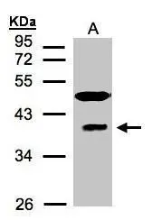
Sample(30 μg of whole cell lysate) A:HeLa S3(GTX14654) 12% SDS PAGE GTX106289 diluted at 1:1500
![RAE1 antibody [C3], C-term detects RAE1 protein at cytoplasm by immunofluorescent analysis. Sample: HeLa cells were fixed in 4% paraformaldehyde at RT for 15 min. Green: RAE1 protein stained by RAE1 antibody [C3], C-term (GTX106289) diluted at 1:500. Blue: Hoechst 33342 staining.](https://www.genetex.com/upload/website/prouct_img/normal/GTX106289/GTX106289_39701_IFA_w_23060120_493.webp "RAE1 antibody [C3], C-term detects RAE1 protein at cytoplasm by immunofluorescent analysis. Sample: HeLa cells were fixed in 4% paraformaldehyde at RT for 15 min. Green: RAE1 protein stained by RAE1 antibody [C3], C-term (GTX106289) diluted at 1:500. Blue: Hoechst 33342 staining.")
Sample(30 μg of whole cell lysate) A:HeLa S3(GTX14654) 12% SDS PAGE GTX106289 diluted at 1:1500
RAE1 antibody [C3], C-term
GTX106289
ApplicationsImmunoFluorescence, Western Blot, ImmunoCytoChemistry
Product group Antibodies
ReactivityHuman
TargetRAE1
Overview
- SupplierGeneTex
- Product NameRAE1 antibody [C3], C-term
- Delivery Days Customer9
- Application Supplier NoteWB: 1:500-1:3000. ICC/IF: 1:100-1:1000. *Optimal dilutions/concentrations should be determined by the researcher.Not tested in other applications.
- ApplicationsImmunoFluorescence, Western Blot, ImmunoCytoChemistry
- CertificationResearch Use Only
- ClonalityPolyclonal
- Concentration1 mg/ml
- ConjugateUnconjugated
- Gene ID8480
- Target nameRAE1
- Target descriptionribonucleic acid export 1
- Target synonymsGle2, MIG14, MRNP41, Mnrp41, dJ481F12.3, dJ800J21.1, mRNA export factor RAE1, RAE1 RNA export 1 homolog, homolog of yeast Rae1 (Bharathi) mRNA-associated protein of 41 kDa (Kraemer), mRNA export factor, mRNA export protein, mRNA-associated protein MRNP 41, mRNA-binding protein, 41-kD, migration-inducing gene 14, rae1 protein homolog
- HostRabbit
- IsotypeIgG
- Protein IDP78406
- Protein NamemRNA export factor RAE1
- Scientific DescriptionMutations in the Schizosaccharomyces pombe Rae1 and Saccharomyces cerevisiae Gle2 genes have been shown to result in accumulation of poly(A)-containing mRNA in the nucleus, suggesting that the encoded proteins are involved in RNA export. The protein encoded by this gene is a homolog of yeast Rae1. It contains four WD40 motifs, and has been shown to localize to distinct foci in the nucleoplasm, to the nuclear rim, and to meshwork-like structures throughout the cytoplasm. This gene is thought to be involved in nucleocytoplasmic transport, and in directly or indirectly attaching cytoplasmic mRNPs to the cytoskeleton. Alternatively spliced transcript variants encoding the same protein have been found for this gene. [provided by RefSeq]
- ReactivityHuman
- Storage Instruction-20°C or -80°C,2°C to 8°C
- UNSPSC41116161
Datasheet
Related products
Product group Antibodies
RAE1 AntibodyCSB-PA019276ESR1HU
ApplicationsWestern Blot, ELISA, ImmunoHistoChemistry
ReactivityHuman, Mouse
TargetRAE1
- SizePrice
Product group Antibodies
Anti-RAE1 AntibodyA31543
ApplicationsImmunoFluorescence, Western Blot, ImmunoHistoChemistry
ReactivityHuman, Mouse, Rat
- SizePrice
Product group Antibodies

Goat anti-RAE1EB06838
ApplicationsWestern Blot, ELISA, ImmunoHistoChemistry
ReactivityBovine, Canine, Human, Mouse, Porcine, Rat
TargetRAE1
- SizePrice

Product group Antibodies
Anti-RAE1 AntibodyHPA048795
ApplicationsImmunoCytoChemistry
ReactivityHuman
TargetRAE1
- SizePrice

Product group Antibodies
Anti-RAE1 Antibody Picoband(r)A04228-2-CARRIER-FREE
ApplicationsFlow Cytometry, ImmunoFluorescence, Western Blot, ELISA, ImmunoCytoChemistry, ImmunoHistoChemistry
ReactivityHuman, Rat
TargetRAE1
- SizePrice
Product group Antibodies

MRNP41 / RAE1 AntibodyLS-C334899
ApplicationsImmunoFluorescence, Western Blot, ImmunoHistoChemistry
ReactivityHuman, Mouse, Rat
TargetRAE1
- SizePrice
Product group Antibodies
RAE1 antibodyGTX33456
ApplicationsImmunoFluorescence, Western Blot, ImmunoCytoChemistry, ImmunoHistoChemistry, ImmunoHistoChemistry Paraffin
ReactivityHuman, Mouse, Rat
TargetRAE1
- SizePrice
Product group Antibodies
RAE1 antibody, C-termGTX89517
ApplicationsWestern Blot, ImmunoHistoChemistry, ImmunoHistoChemistry Paraffin
ReactivityHuman
TargetRAE1
- SizePrice
Product group Antibodies
Anti-RAE1 Antibody144-06713
ApplicationsImmunoFluorescence, Western Blot, ImmunoHistoChemistry
ReactivityHuman, Mouse, Rat
TargetRAE1
- SizePrice