

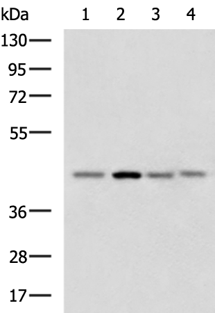
Gel: 8%SDS-PAGE Lysate: 40 microg Lane 1-4: HepG2 Jurkat cell Mouse brain tissue Human cerebrum tissue lysates Primary antibody: TA364955 (RAE1 Antibody) at dilution 1/1000 Secondary antibody: Goat anti rabbit IgG at 1/5000 dilution Exposure time: 1 minute
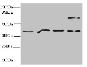
Gel: 8%SDS-PAGE Lysate: 40 microg Lane 1-4: HepG2 Jurkat cell Mouse brain tissue Human cerebrum tissue lysates Primary antibody: TA364955 (RAE1 Antibody) at dilution 1/1000 Secondary antibody: Goat anti rabbit IgG at 1/5000 dilution Exposure time: 1 minute
RAE1 Rabbit Polyclonal Antibody
TA364955
Overview
- SupplierOriGene
- Product NameRAE1 Rabbit Polyclonal Antibody
- Delivery Days Customer14
- ApplicationsWestern Blot, ImmunoHistoChemistry
- CertificationResearch Use Only
- ClonalityPolyclonal
- Gene ID8480
- Target nameRAE1
- Target descriptionribonucleic acid export 1
- Target synonymsdJ481F12.3; dJ800J21.1; Gle2; homolog of yeast Rae1 (Bharathi) mRNA-associated protein of 41 kDa (Kraemer); MIG14; migration-inducing gene 14; Mnrp41; mRNA export factor; mRNA export protein; mRNA-associated protein MRNP 41; mRNA-binding protein, 41-kD; MRNP41; rae1 protein homolog; RAE1 RNA export 1 homolog
- HostRabbit
- IsotypeIgG
- Protein IDP78406
- Protein NamemRNA export factor
- Scientific DescriptionRAE1 rabbit polyclonal antibody
- Storage Instruction-20°C
- UNSPSC12352203
Related products
Product group Antibodies
Anti-RAE1 Antibody144-06713
ApplicationsImmunoFluorescence, Western Blot, ImmunoHistoChemistry
TargetRAE1
- SizePrice
Product group Antibodies
RAE1 AntibodyCSB-PA019276ESR1HU
ApplicationsWestern Blot, ELISA, ImmunoHistoChemistry
ReactivityHuman, Mouse
TargetRAE1
- SizePrice
Product group Antibodies

MRNP41 / RAE1 AntibodyLS-C334899
ApplicationsImmunoFluorescence, Western Blot, ImmunoHistoChemistry
TargetRAE1
- SizePrice
Product group Antibodies
RAE1 Polyclonal AntibodyBS-3791R
ApplicationsImmunoFluorescence, ELISA, ImmunoCytoChemistry, ImmunoHistoChemistry, ImmunoHistoChemistry Frozen, ImmunoHistoChemistry Paraffin
TargetRAE1
- SizePrice

Product group Antibodies
Anti-RAE1 AntibodyHPA048795
ApplicationsImmunoCytoChemistry
ReactivityHuman
TargetRAE1
- SizePrice

Product group Antibodies
Anti-RAE1 Antibody Picoband(r)A04228-2-CARRIER-FREE
ApplicationsFlow Cytometry, ImmunoFluorescence, Western Blot, ELISA, ImmunoCytoChemistry, ImmunoHistoChemistry
TargetRAE1
- SizePrice
Product group Antibodies
RAE1 antibody [C3], C-termGTX106289
ApplicationsImmunoFluorescence, Western Blot, ImmunoCytoChemistry
TargetRAE1
- SizePrice
Product group Antibodies

Goat anti-RAE1EB06838
ApplicationsWestern Blot, ELISA, ImmunoHistoChemistry
TargetRAE1
- SizePrice
Product group Antibodies
Anti-RAE1 AntibodyA31543
ApplicationsImmunoFluorescence, Western Blot, ImmunoHistoChemistry
- SizePrice