

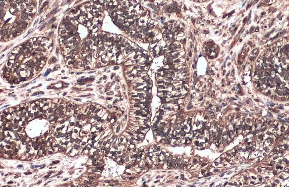
Raf1 antibody [N3C3] detects Raf1 protein at cell membrane and cytoplasm by immunohistochemical analysis. Sample: Paraffin-embedded human endometrial carcinoma. Raf1 stained by Raf1 antibody [N3C3] (GTX107763) diluted at 1:500. Antigen Retrieval: Citrate buffer, pH 6.0, 15 min
 antibody at 1:200 dilution.")
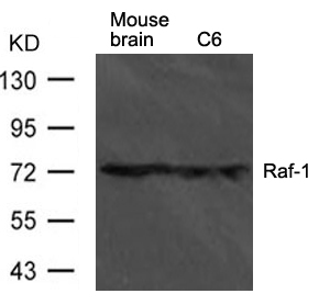
![Non-transfected (–) and transfected (+) 293T whole cell extracts (30 μg) were separated by 7.5% SDS-PAGE, and the membrane was blotted with Raf1 antibody [N3C3] (GTX107763) diluted at 1:1000. The HRP-conjugated anti-rabbit IgG antibody (GTX213110-01) was used to detect the primary antibody.](https://www.genetex.com/upload/website/prouct_img/normal/GTX107763/GTX107763_42907_20181109_WB_B_w_23060120_748.webp "Non-transfected (–) and transfected (+) 293T whole cell extracts (30 μg) were separated by 7.5% SDS-PAGE, and the membrane was blotted with Raf1 antibody [N3C3] (GTX107763) diluted at 1:1000. The HRP-conjugated anti-rabbit IgG antibody (GTX213110-01) was used to detect the primary antibody.")
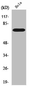
![Untreated (–) and treated (+) HeLa whole cell extracts (30 μg) were separated by 7.5% SDS-PAGE, and the membrane was blotted with Raf1 antibody [N3C3] (GTX107763) diluted at 1:1000. The HRP-conjugated anti-rabbit IgG antibody (GTX213110-01) was used to detect the primary antibody.](https://www.genetex.com/upload/website/prouct_img/normal/GTX107763/GTX107763_43602_20190524_WB_treatment_PMA_w_23060120_686.webp "Untreated (–) and treated (+) HeLa whole cell extracts (30 μg) were separated by 7.5% SDS-PAGE, and the membrane was blotted with Raf1 antibody [N3C3] (GTX107763) diluted at 1:1000. The HRP-conjugated anti-rabbit IgG antibody (GTX213110-01) was used to detect the primary antibody.")
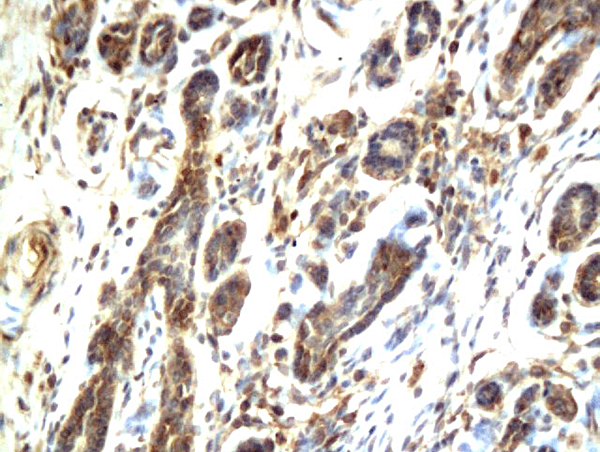
![Raf1 antibody [N3C3] detects Raf1 protein at nucleus on mouse muscle by immunohistochemical analysis. Sample: Paraffin-embedded mouse muscle. Raf1 antibody [N3C3] (GTX107763) dilution: 1:500.
Antigen Retrieval: Trilogy? (EDTA based, pH 8.0) buffer, 15min](https://www.genetex.com/upload/website/prouct_img/normal/GTX107763/GTX107763_40016_IHC_M_w_23060120_339.webp "Raf1 antibody [N3C3] detects Raf1 protein at nucleus on mouse muscle by immunohistochemical analysis. Sample: Paraffin-embedded mouse muscle. Raf1 antibody [N3C3] (GTX107763) dilution: 1:500.
Antigen Retrieval: Trilogy? (EDTA based, pH 8.0) buffer, 15min")
Raf1 antibody [N3C3] detects Raf1 protein at cell membrane and cytoplasm by immunohistochemical analysis. Sample: Paraffin-embedded human endometrial carcinoma. Raf1 stained by Raf1 antibody [N3C3] (GTX107763) diluted at 1:500. Antigen Retrieval: Citrate buffer, pH 6.0, 15 min
Raf1 antibody [N3C3]
GTX107763
ApplicationsImmunoFluorescence, Western Blot, ImmunoCytoChemistry, ImmunoHistoChemistry, ImmunoHistoChemistry Paraffin
Product group Antibodies
ReactivityHuman, Mouse
TargetRAF1
Overview
- SupplierGeneTex
- Product NameRaf1 antibody [N3C3]
- Delivery Days Customer9
- Application Supplier NoteWB: 1:500-1:3000. ICC/IF: 1:100-1:1000. IHC-P: 1:100-1:1000. *Optimal dilutions/concentrations should be determined by the researcher.Not tested in other applications.
- ApplicationsImmunoFluorescence, Western Blot, ImmunoCytoChemistry, ImmunoHistoChemistry, ImmunoHistoChemistry Paraffin
- CertificationResearch Use Only
- ClonalityPolyclonal
- Concentration0.36 mg/ml
- ConjugateUnconjugated
- Gene ID5894
- Target nameRAF1
- Target descriptionRaf-1 proto-oncogene, serine/threonine kinase
- Target synonymsCMD1NN, CRAF, NS5, Raf-1, c-Raf, RAF proto-oncogene serine/threonine-protein kinase, C-Raf proto-oncogene, serine/threonine kinase, Oncogene RAF1, proto-oncogene c-RAF, raf proto-oncogene serine/threonine protein kinase, v-raf-1 murine leukemia viral oncogene homolog 1, v-raf-1 murine leukemia viral oncogene-like protein 1
- HostRabbit
- IsotypeIgG
- Protein IDP04049
- Protein NameRAF proto-oncogene serine/threonine-protein kinase
- Scientific DescriptionThis gene is the cellular homolog of viral raf gene (v-raf). The encoded protein is a MAP kinase kinase kinase (MAP3K), which functions downstream of the Ras family of membrane associated GTPases to which it binds directly. Once activated, the cellular RAF1 protein can phosphorylate to activate the dual specificity protein kinases MEK1 and MEK2, which in turn phosphorylate to activate the serine/threonine specific protein kinases, ERK1 and ERK2. Activated ERKs are pleiotropic effectors of cell physiology and play an important role in the control of gene expression involved in the cell division cycle, apoptosis, cell differentiation and cell migration. Mutations in this gene are associated with Noonan syndrome 5 and LEOPARD syndrome 2. [provided by RefSeq]
- ReactivityHuman, Mouse
- Storage Instruction-20°C or -80°C,2°C to 8°C
- UNSPSC41116161
Datasheet
Related products
Product group Antibodies

C-RAF (Phospho-Tyr341) AntibodyABX012449
ApplicationsWestern Blot, ELISA, ImmunoHistoChemistry
- SizePrice
Product group Antibodies
Anti-RAF1 Antibody144-00223
ApplicationsImmunoFluorescence, Western Blot, ImmunoHistoChemistry
ReactivityHuman, Mouse, Rat
TargetRAF1
- SizePrice
Product group Antibodies
Anti-Raf1 AntibodyA42606
ApplicationsWestern Blot
ReactivityHuman, Mouse, Rat
- SizePrice

Product group Antibodies
Anti-Raf1 Antibody Picoband(r)A00446-1-CARRIER-FREE
ApplicationsFlow Cytometry, Western Blot
ReactivityHuman, Mouse, Rat
TargetRAF1
- SizePrice
Product group Antibodies
RAF1 AntibodyCSB-PA003921
ApplicationsImmunoFluorescence, Western Blot, ELISA, ImmunoHistoChemistry
ReactivityHuman, Mouse, Rat
TargetRAF1
- SizePrice
Product group Antibodies
RAF1 Polyclonal AntibodyCAC13895
ApplicationsImmunoFluorescence, ImmunoPrecipitation, Western Blot, ELISA, ImmunoHistoChemistry
TargetRAF1
- SizePrice
Product group Antibodies
RAF1 Polyclonal AntibodyBS-1703R
ApplicationsImmunoFluorescence, ELISA, ImmunoCytoChemistry, ImmunoHistoChemistry, ImmunoHistoChemistry Frozen, ImmunoHistoChemistry Paraffin
ReactivityBovine, Canine, Equine, Human, Mouse, Rabbit, Rat
TargetRAF1
- SizePrice
Product group Antibodies
Raf1 antibody [410]GTX20656
ApplicationsWestern Blot, ELISA
ReactivityHuman
TargetRAF1
- SizePrice
Product group Antibodies
ApplicationsWestern Blot, ImmunoHistoChemistry
ReactivityHuman, Mouse, Rat
TargetRAF1
- SizePrice