

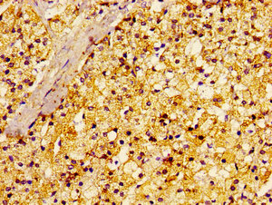
Immunohistochemistry of paraffin-embedded human adrenal gland tissue using CSB-PA019296LA01HU at dilution of 1:100
.")
Immunohistochemistry of paraffin-embedded human adrenal gland tissue using CSB-PA019296LA01HU at dilution of 1:100
RALA Antibody
CSB-PA019296LA01HU
ApplicationsImmunoFluorescence, ELISA, ImmunoHistoChemistry
Product group Antibodies
ReactivityHuman
TargetRALA
Overview
- SupplierCusabio
- Product NameRALA Antibody
- Delivery Days Customer20
- ApplicationsImmunoFluorescence, ELISA, ImmunoHistoChemistry
- CertificationResearch Use Only
- ClonalityPolyclonal
- ConjugateUnconjugated
- Gene ID5898
- Target nameRALA
- Target descriptionRAS like proto-oncogene A
- Target synonymsHINCONS, RAL, ras-related protein Ral-A, RALA Ras like proto-oncogene A, RAS-like protein A, Ras family small GTP binding protein RALA, ras related GTP binding protein A, v-ral simian leukemia viral oncogene homolog A (ras related)
- HostRabbit
- IsotypeIgG
- Protein IDP11233
- Protein NameRas-related protein Ral-A
- Scientific DescriptionMultifunctional GTPase involved in a variety of cellular processes including gene expression, cell migration, cell proliferation, oncogenic transformation and membrane trafficking. Accomplishes its multiple functions by interacting with distinct downstream effectors. Acts as a GTP sensor for GTP-dependent exocytosis of dense core vesicles. Plays a role in the early stages of cytokinesis and is required to tether the exocyst to the cytokinetic furrow. The RALA-exocyst complex regulates integrin-dependent membrane raft exocytosis and growth signaling. Key regulator of LPAR1 signaling and competes with GRK2 for binding to LPAR1 thus affecting the signaling properties of the receptor. Required for anchorage-independent proliferation of transformed cells.
- ReactivityHuman
- Storage Instruction-20°C or -80°C
- UNSPSC41116161
Related products
Product group Antibodies
Anti-RALA AntibodyA17135
ApplicationsImmunoFluorescence, Western Blot, ImmunoCytoChemistry
ReactivityMouse, Rat
- SizePrice
Product group Antibodies
Anti-Mouse/Rat RALA Antibody144-00541
ApplicationsImmunoFluorescence, Western Blot
ReactivityHuman, Mouse, Rat
TargetRALA
- SizePrice
Product group Antibodies

RALA / RAL AntibodyLS-C747108
ApplicationsWestern Blot
ReactivityHuman, Mouse, Rat
TargetRALA
- SizePrice
Product group Antibodies
ApplicationsImmunoPrecipitation, Western Blot, ImmunoCytoChemistry, ImmunoHistoChemistry
ReactivityMouse, Rat
TargetRALA
- SizePrice

Product group Antibodies
Anti-RALA AntibodyHPA065232
ApplicationsWestern Blot, ImmunoHistoChemistry
ReactivityHuman
TargetRALA
- SizePrice
Product group Antibodies
RALA antibodyGTX114204
ApplicationsWestern Blot
ReactivityAmphibian, Human, Mammals, Mouse, Rat
TargetRALA
- SizePrice
Product group Antibodies
Anti-RALA AntibodyCAB0541
ApplicationsWestern Blot, ELISA
ReactivityMouse
TargetRALA
- SizePrice

Product group Antibodies
Anti-RALA Antibody Picoband(r)PB9794-CARRIER-FREE
ApplicationsWestern Blot
ReactivityBovine, Canine, Chicken, Equine, Human, Monkey, Mouse, Rabbit, Rat, Zebra Fish
TargetRALA
- SizePrice