

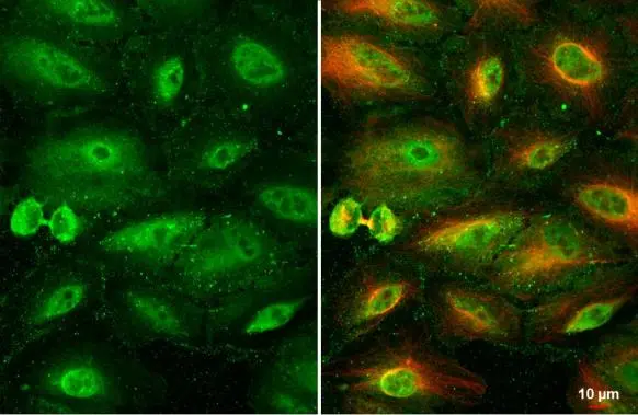
RBPMS antibody detects RBPMS protein at cytoplasm and nucleus by immunofluorescent analysis. Sample: A549 cells were fixed in 4% paraformaldehyde at RT for 15 min. Green: RBPMS stained by RBPMS antibody (GTX118619) diluted at 1:500. Red: alpha Tubulin, a cytoskeleton marker, stained by alpha Tubulin antibody [GT114] (GTX628802) diluted at 1:1000.
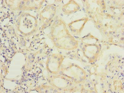
 diluted at 1:500. Antigen Retrieval: Citrate buffer, pH 6.0, 15 min")
 diluted at 1:2000. Antigen Retrieval: Citrate buffer, pH 6.0, 15 min")
 diluted at 1:500. Antigen Retrieval: Citrate buffer, pH 6.0, 15 min")
![RBPMS antibody detects RBPMS protein by immunohistochemical analysis. Samples: Paraffin-embedded mouse retina. Green: RBPMS protein stained by RBPMS antibody (GTX118619) diluted at 1:250. Red: beta Tubulin 3/ Tuj1, a marker, stained by beta Tubulin 3/ Tuj1 antibody [GT1338] (GTX631831) diluted at 1:500. Blue: Fluoroshield with DAPI (GTX30920).
Antigen Retrieval: Citrate buffer, pH 6.0, 15 min](https://www.genetex.com/upload/website/prouct_img/normal/GTX118619/GTX118619_42810_20171127_IHC-P_M_w_23060519_346.webp "RBPMS antibody detects RBPMS protein by immunohistochemical analysis. Samples: Paraffin-embedded mouse retina. Green: RBPMS protein stained by RBPMS antibody (GTX118619) diluted at 1:250. Red: beta Tubulin 3/ Tuj1, a marker, stained by beta Tubulin 3/ Tuj1 antibody [GT1338] (GTX631831) diluted at 1:500. Blue: Fluoroshield with DAPI (GTX30920).
Antigen Retrieval: Citrate buffer, pH 6.0, 15 min")
 diluted at 1:2000. Antigen Retrieval: Citrate buffer, pH 6.0, 15 min")
 and transfected (+) 293T whole cell extracts (30 μg) were separated by 12% SDS-PAGE, and the membrane was blotted with RBPMS antibody (GTX118619) diluted at 1:1000. The HRP-conjugated anti-rabbit IgG antibody (GTX213110-01) was used to detect the primary antibody.")
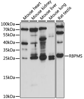
 were separated by 12% SDS-PAGE, and the membrane was blotted with RBPMS antibody (GTX118619) diluted at 1:500. The HRP-conjugated anti-rabbit IgG antibody (GTX213110-01) was used to detect the primary antibody.")
 and transfected (+) HCT-116 whole cell extract were separated by 12% SDS-PAGE, and the membrane was blotted with RBPMS antibody (GTX118619) diluted at 1:500. The HRP-conjugated anti-rabbit IgG antibody (GTX213110-01) was used to detect the primary antibody.")
![RBPMS antibody detects RBPMS protein at cytoplasm and nucleus by immunofluorescent analysis. Sample: A431 cells were fixed in 4% paraformaldehyde at RT for 15 min. Green: RBPMS stained by RBPMS antibody (GTX118619) diluted at 1:500. Red: alpha Tubulin, a cytoskeleton marker, stained by alpha Tubulin antibody [GT114] (GTX628802) diluted at 1:1000. Blue: Fluoroshield with DAPI (GTX30920).](https://www.genetex.com/upload/website/prouct_img/normal/GTX118619/GTX118619_45260_20240329_ICC_IF_24041019_824.webp "RBPMS antibody detects RBPMS protein at cytoplasm and nucleus by immunofluorescent analysis. Sample: A431 cells were fixed in 4% paraformaldehyde at RT for 15 min. Green: RBPMS stained by RBPMS antibody (GTX118619) diluted at 1:500. Red: alpha Tubulin, a cytoskeleton marker, stained by alpha Tubulin antibody [GT114] (GTX628802) diluted at 1:1000. Blue: Fluoroshield with DAPI (GTX30920).")
RBPMS antibody detects RBPMS protein at cytoplasm and nucleus by immunofluorescent analysis. Sample: A549 cells were fixed in 4% paraformaldehyde at RT for 15 min. Green: RBPMS stained by RBPMS antibody (GTX118619) diluted at 1:500. Red: alpha Tubulin, a cytoskeleton marker, stained by alpha Tubulin antibody [GT114] (GTX628802) diluted at 1:1000.
RBPMS antibody
GTX118619
ApplicationsImmunoFluorescence, Western Blot, ImmunoCytoChemistry, ImmunoHistoChemistry, ImmunoHistoChemistry Paraffin
Product group Antibodies
ReactivityHuman, Monkey, Mouse, Rat
TargetRBPMS
Overview
- SupplierGeneTex
- Product NameRBPMS antibody
- Delivery Days Customer9
- Application Supplier NoteWB: 1:500-1:3000. ICC/IF: 1:100-1:1000. IHC-P: 1:100-1:1000. *Optimal dilutions/concentrations should be determined by the researcher.Not tested in other applications.
- ApplicationsImmunoFluorescence, Western Blot, ImmunoCytoChemistry, ImmunoHistoChemistry, ImmunoHistoChemistry Paraffin
- CertificationResearch Use Only
- ClonalityPolyclonal
- Concentration1.41 mg/ml
- ConjugateUnconjugated
- Gene ID11030
- Target nameRBPMS
- Target descriptionRNA binding protein, mRNA processing factor
- Target synonymsHERMES, RNA-binding protein with multiple splicing, RNA binding protein with multiple splicing, heart and RRM expressed sequence
- HostRabbit
- IsotypeIgG
- Protein IDQ93062
- Protein NameRNA-binding protein with multiple splicing
- Scientific DescriptionThis gene encodes a member of the RRM family of RNA-binding proteins. The RRM domain is between 80-100 amino acids in length and family members contain one to four copies of the domain. The RRM domain consists of two short stretches of conserved sequence called RNP1 and RNP2, as well as a few highly conserved hydrophobic residues. The protein encoded by this gene has a single, putative RRM domain in its N-terminus. Alternative splicing results in multiple transcript variants encoding different isoforms. [provided by RefSeq]
- ReactivityHuman, Monkey, Mouse, Rat
- Storage Instruction-20°C or -80°C,2°C to 8°C
- UNSPSC41116161
Datasheet
Related products
Product group Antibodies
Anti-RBPMS AntibodyA88779
ApplicationsWestern Blot
ReactivityHuman, Mouse, Rat
- SizePrice

Product group Antibodies
Anti-RBPMS Antibody Picoband(r)A07130-2-CARRIER-FREE
ApplicationsFlow Cytometry, Western Blot, ELISA, ImmunoHistoChemistry
ReactivityHuman, Mouse, Rat
TargetRBPMS
- SizePrice
Product group Antibodies
Anti-RBPMS Antibody144-63730
ApplicationsImmunoFluorescence, Western Blot
ReactivityHuman, Mouse, Rat
TargetRBPMS
- SizePrice
Product group Antibodies

RBPMS / Hermes AntibodyLS-C750367
ApplicationsWestern Blot
ReactivityHuman, Mouse, Rat
TargetRBPMS
- SizePrice
Product group Antibodies
RBPMS AntibodyCSB-PA04569A0RB
ApplicationsImmunoFluorescence, ELISA, ImmunoHistoChemistry
ReactivityHuman
TargetRBPMS
- SizePrice
Product group Antibodies
Anti-RBPMS AntibodyHPA056999
ApplicationsWestern Blot
ReactivityHuman
TargetRBPMS
- SizePrice
![RBPMS antibody [HL3090] detects RBPMS protein by immunohistochemical analysis. Sample: Mouse whole-mount retina were fixed in 4% paraformaldehyde at RT for 30 min. Green: RBPMS stained by RBPMS antibody [HL3090] (GTX640536) diluted at 1:1000. Red: beta Tubulin 3/ Tuj1, stained by beta Tubulin 3/ Tuj1 antibody (GTX631836) diluted at 1:500. Blue: Hoechst 33342 staining. 4% paraformaldehyde at RT for 30 min](https://www.genetex.com/upload/website/prouct_img/normal/GTX640536/GTX640536_T-45453_19010514_IHC-P_M_24111918_696.webp)
Product group Antibodies
RBPMS antibody [HL3090]GTX640536
ApplicationsImmunoHistoChemistry, ImmunoHistoChemistry Frozen, ImmunoHistoChemistry Paraffin
ReactivityHuman, Mouse
TargetRBPMS
- SizePrice
![RBPMS antibody [HL1105] detects RBPMS protein by immunohistochemical analysis. Sample: Paraffin-embedded mouse tissues. RBPMS stained by RBPMS antibody [HL1105] (GTX636320) diluted at 1:100. Antigen Retrieval: Citrate buffer, pH 6.0, 15 min](https://www.genetex.com/upload/website/prouct_img/normal/GTX636320/GTX636320_44620_20221227_IHC-P_multiple_M_22122821_891.webp)
Product group Antibodies
RBPMS antibody [HL1105]GTX636320
ApplicationsWestern Blot, ImmunoHistoChemistry, ImmunoHistoChemistry Paraffin
ReactivityHuman, Mouse, Rat
TargetRBPMS
- SizePrice
![RBPMS antibody [HL1106] detects RBPMS protein by immunohistochemical analysis. Sample: Paraffin-embedded mouse tissues. RBPMS stained by RBPMS antibody [HL1106] (GTX636321) diluted at 1:100. Antigen Retrieval: Citrate buffer, pH 6.0, 15 min](https://www.genetex.com/upload/website/prouct_img/normal/GTX636321/GTX636321_44620_20221227_IHC-P_multiple_M_22122821_217.webp)
Product group Antibodies
RBPMS antibody [HL1106]GTX636321
ApplicationsWestern Blot, ImmunoHistoChemistry, ImmunoHistoChemistry Paraffin
ReactivityHuman, Mouse, Rat
TargetRBPMS
- SizePrice