

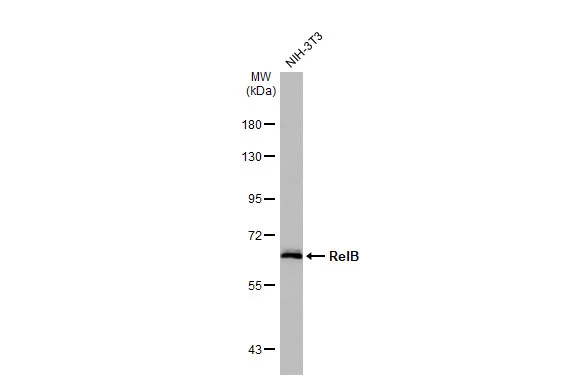
Whole cell extract (30 μg) was separated by 7.5% SDS-PAGE, and the membrane was blotted with RelB antibody [HL2222] (GTX638267) diluted at 1:1000. The HRP-conjugated anti-rabbit IgG antibody (GTX213110-01) was used to detect the primary antibody.
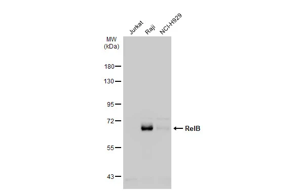
![Various whole cell extracts (30 μg) were separated by 7.5% SDS-PAGE, and the membrane was blotted with RelB antibody [HL2222] (GTX638267) diluted at 1:1000. The HRP-conjugated anti-rabbit IgG antibody (GTX213110-01) was used to detect the primary antibody, and the signal was developed with Trident ECL plus-Enhanced.](https://www.genetex.com/upload/website/prouct_img/normal/GTX638267/GTX638267_T-44956_20230217_WB_R_23022022_600.webp "Various whole cell extracts (30 μg) were separated by 7.5% SDS-PAGE, and the membrane was blotted with RelB antibody [HL2222] (GTX638267) diluted at 1:1000. The HRP-conjugated anti-rabbit IgG antibody (GTX213110-01) was used to detect the primary antibody, and the signal was developed with Trident ECL plus-Enhanced.")
![RelB antibody [HL2222] detects RelB protein at cytoplasm and nucleus by immunohistochemical analysis. Sample: Paraffin-embedded rat colon. RelB stained by RelB antibody [HL2222] (GTX638267) diluted at 1:200. Antigen Retrieval: Citrate buffer, pH 6.0, 15 min](https://www.genetex.com/upload/website/prouct_img/normal/GTX638267/GTX638267_T-44956_20230325_IHC-P_R_23032819_282.webp "RelB antibody [HL2222] detects RelB protein at cytoplasm and nucleus by immunohistochemical analysis. Sample: Paraffin-embedded rat colon. RelB stained by RelB antibody [HL2222] (GTX638267) diluted at 1:200. Antigen Retrieval: Citrate buffer, pH 6.0, 15 min")
![Various whole cell extracts (30 μg) were separated by 7.5% SDS-PAGE, and the membrane was blotted with RelB antibody [HL2222] (GTX638267) diluted at 1:1000. The HRP-conjugated anti-rabbit IgG antibody (GTX213110-01) was used to detect the primary antibody. Corresponding RNA expression data for the same cell lines are based on Human Protein Atlas program.](https://www.genetex.com/upload/website/prouct_img/normal/GTX638267/GTX638267_45026_20230428_WB_TPM_watermark_23050223_563.webp "Various whole cell extracts (30 μg) were separated by 7.5% SDS-PAGE, and the membrane was blotted with RelB antibody [HL2222] (GTX638267) diluted at 1:1000. The HRP-conjugated anti-rabbit IgG antibody (GTX213110-01) was used to detect the primary antibody. Corresponding RNA expression data for the same cell lines are based on Human Protein Atlas program.")
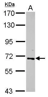
![Non-transfected (–) and transfected (+) 293T whole cell extracts (30 μg) were separated by 7.5% SDS-PAGE, and the membrane was blotted with RelB antibody [HL2222] (GTX638267) diluted at 1:5000. The HRP-conjugated anti-rabbit IgG antibody (GTX213110-01) was used to detect the primary antibody.](https://www.genetex.com/upload/website/prouct_img/normal/GTX638267/GTX638267_45026_20230707_WB_B_23071223_371.webp "Non-transfected (–) and transfected (+) 293T whole cell extracts (30 μg) were separated by 7.5% SDS-PAGE, and the membrane was blotted with RelB antibody [HL2222] (GTX638267) diluted at 1:5000. The HRP-conjugated anti-rabbit IgG antibody (GTX213110-01) was used to detect the primary antibody.")
![Immunoprecipitation of RelB protein from Raji whole cell extract using 5 μg of RelB antibody [HL2222] (GTX638267). Western blot analysis was performed using RelB antibody [HL2222] (GTX638267). EasyBlot HRP-conjugated anti rabbit IgG antibody (GTX221666-01).](https://www.genetex.com/upload/website/prouct_img/normal/GTX638267/GTX638267_T-44956_20240419_IP_24042400_827.webp "Immunoprecipitation of RelB protein from Raji whole cell extract using 5 μg of RelB antibody [HL2222] (GTX638267). Western blot analysis was performed using RelB antibody [HL2222] (GTX638267). EasyBlot HRP-conjugated anti rabbit IgG antibody (GTX221666-01).")
Whole cell extract (30 μg) was separated by 7.5% SDS-PAGE, and the membrane was blotted with RelB antibody [HL2222] (GTX638267) diluted at 1:1000. The HRP-conjugated anti-rabbit IgG antibody (GTX213110-01) was used to detect the primary antibody.
RelB antibody [HL2222]
GTX638267
ApplicationsImmunoPrecipitation, Western Blot, ImmunoHistoChemistry, ImmunoHistoChemistry Paraffin
Product group Antibodies
ReactivityHuman, Mouse, Rat
TargetRELB
Overview
- SupplierGeneTex
- Product NameRelB antibody [HL2222]
- Delivery Days Customer9
- Application Supplier NoteWB: 1:500-1:3000. *Optimal dilutions/concentrations should be determined by the researcher.Not tested in other applications.
- ApplicationsImmunoPrecipitation, Western Blot, ImmunoHistoChemistry, ImmunoHistoChemistry Paraffin
- CertificationResearch Use Only
- ClonalityMonoclonal
- Clone IDHL2222
- Concentration2 mg/ml
- ConjugateUnconjugated
- Gene ID5971
- Target nameRELB
- Target descriptionRELB proto-oncogene, NF-kB subunit
- Target synonymsI-REL, IMD53, IREL, REL-B, transcription factor RelB, v-rel avian reticuloendotheliosis viral oncogene homolog B (nuclear factor of kappa light polypeptide gene enhancer in B-cells 3), v-rel reticuloendotheliosis viral oncogene homolog B, nuclear factor of kappa light polypeptide gene enhancer in B-cells 3
- HostRabbit
- IsotypeIgG
- Protein IDQ01201
- Protein NameTranscription factor RelB
- Scientific DescriptionEnables mRNA N6-methyladenosine dioxygenase activity. Involved in RNA metabolic process; mRNA export from nucleus; and response to hypoxia. Located in Golgi apparatus; cytosol; and nuclear speck. [provided by Alliance of Genome Resources, Apr 2022]
- ReactivityHuman, Mouse, Rat
- Storage Instruction-20°C or -80°C,2°C to 8°C
- UNSPSC41116161
Datasheet
Related products

Product group Antibodies
ApplicationsWestern Blot
ReactivityHuman, Mouse
- SizePrice
Product group Antibodies
Anti-RELB Antibody144-00519
ApplicationsWestern Blot
ReactivityHuman, Mouse
TargetRELB
- SizePrice
Product group Antibodies
ApplicationsFlow Cytometry, Western Blot, ImmunoCytoChemistry
ReactivityHuman, Mouse, Rat
TargetRELB
- SizePrice
Product group Antibodies
Phospho-RELB (S552) AntibodyCSB-PA010548
ApplicationsWestern Blot, ELISA, ImmunoHistoChemistry
ReactivityHuman, Mouse
TargetRELB
- SizePrice
Product group Antibodies
RELB Polyclonal AntibodyCAC15022
ApplicationsImmunoFluorescence, Western Blot, ELISA
ReactivityMouse
TargetRELB
- SizePrice
Product group Antibodies
RelB antibodyGTX102332
ApplicationsWestern Blot
ReactivityHuman
TargetRELB
- SizePrice
Product group Antibodies
RelB antibodyGTX102333
ApplicationsImmunoPrecipitation, Western Blot, ChIP Chromatin ImmunoPrecipitation, ImmunoHistoChemistry, ImmunoHistoChemistry Paraffin
ReactivityHuman, Mouse
TargetRELB
- SizePrice
Product group Antibodies

RELB Antibody (C-Terminus)LS-C368628
ApplicationsWestern Blot, ImmunoHistoChemistry, ImmunoHistoChemistry Paraffin
ReactivityHuman, Mouse, Rat
TargetRELB
- SizePrice
Product group Antibodies
RelB (phospho Ser552) antibodyGTX78951
ApplicationsWestern Blot, ImmunoHistoChemistry, ImmunoHistoChemistry Paraffin
ReactivityHuman
TargetRELB
- SizePrice

Product group Antibodies
Anti-RELB AntibodyHPA040506
ApplicationsImmunoCytoChemistry, ImmunoHistoChemistry
ReactivityHuman
TargetRELB
- SizePrice