

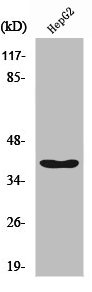
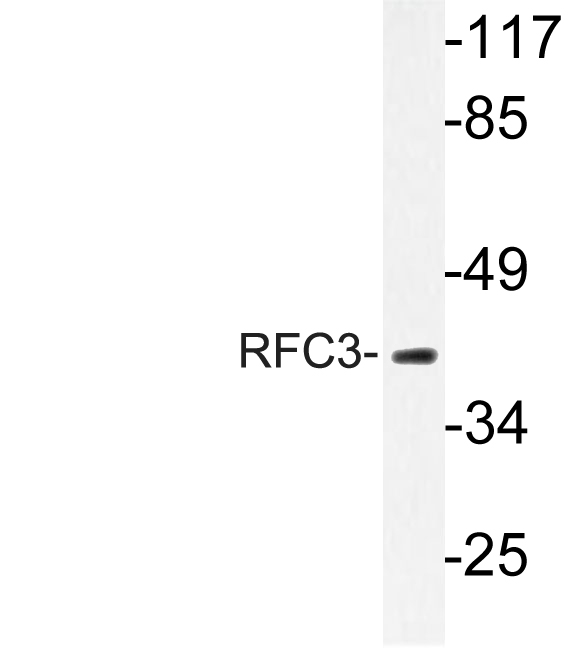
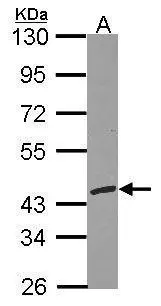
Western Blot analysis of HepG2 cells using RFC3 Polyclonal Antibody
Western Blot analysis of HepG2 cells using RFC3 Polyclonal Antibody
RFC3 Antibody
CSB-PA003964
ApplicationsWestern Blot, ELISA, ImmunoHistoChemistry
Product group Antibodies
ReactivityHuman, Mouse, Rat
TargetRFC3
Overview
- SupplierCusabio
- Product NameRFC3 Antibody
- Delivery Days Customer20
- ApplicationsWestern Blot, ELISA, ImmunoHistoChemistry
- CertificationResearch Use Only
- ClonalityPolyclonal
- ConjugateUnconjugated
- Gene ID5983
- Target nameRFC3
- Target descriptionreplication factor C subunit 3
- Target synonymsRFC38, replication factor C subunit 3, A1 38 kDa subunit, RF-C 38 kDa subunit, RFC, 38 kD subunit, activator 1 38 kDa subunit, activator 1 subunit 3, replication factor C (activator 1) 3, 38kDa, replication factor C 38 kDa subunit
- HostRabbit
- IsotypeIgG
- Protein IDP40938
- Protein NameReplication factor C subunit 3
- ReactivityHuman, Mouse, Rat
- Storage Instruction-20°C or -80°C
- UNSPSC41116161
Related products
Product group Antibodies
Anti-RFC3 AntibodyA96133
ApplicationsWestern Blot, ELISA, ImmunoHistoChemistry
ReactivityHuman, Mouse, Rat
- SizePrice

Product group Antibodies
Anti-RFC3 Antibody Picoband(r)A06442-2-CARRIER-FREE
ApplicationsFlow Cytometry, ImmunoFluorescence, Western Blot, ELISA, ImmunoCytoChemistry
ReactivityHuman, Mouse, Rat
TargetRFC3
- SizePrice

Product group Antibodies
Anti-RFC3 AntibodyHPA030149
ApplicationsWestern Blot, ImmunoCytoChemistry, ImmunoHistoChemistry
ReactivityHuman
TargetRFC3
- SizePrice
Product group Antibodies

RFC3 Antibody (aa80-110)LS-C287111
ApplicationsWestern Blot, ImmunoHistoChemistry
ReactivityHuman
TargetRFC3
- SizePrice
Product group Antibodies
RFC3 antibodyGTX103706
ApplicationsImmunoFluorescence, Western Blot, ImmunoCytoChemistry, ImmunoHistoChemistry, ImmunoHistoChemistry Paraffin
ReactivityHuman
TargetRFC3
- SizePrice
Product group Antibodies
Anti-RFC3 Antibody144-04075
ApplicationsWestern Blot
ReactivityHuman, Mouse, Rat
TargetRFC3
- SizePrice