

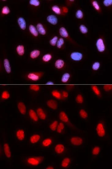
ICC/IF analysis of U2OS cells using GTX33465 RFC4 antibody. Blue : DAPI


ICC/IF analysis of U2OS cells using GTX33465 RFC4 antibody. Blue : DAPI
RFC4 antibody
GTX33465
ApplicationsImmunoFluorescence, ImmunoPrecipitation, Western Blot, ImmunoCytoChemistry
Product group Antibodies
ReactivityHuman
TargetRFC4
Overview
- SupplierGeneTex
- Product NameRFC4 antibody
- Delivery Days Customer9
- Application Supplier NoteWB: 1:500 - 1:2000. ICC/IF: 1:50 - 1:100. IP: 1:50 - 1:200. *Optimal dilutions/concentrations should be determined by the researcher.Not tested in other applications.
- ApplicationsImmunoFluorescence, ImmunoPrecipitation, Western Blot, ImmunoCytoChemistry
- CertificationResearch Use Only
- ClonalityPolyclonal
- ConjugateUnconjugated
- Gene ID5984
- Target nameRFC4
- Target descriptionreplication factor C subunit 4
- Target synonymsA1, MRMNS, RFC37, replication factor C subunit 4, A1 37 kDa subunit, RF-C 37 kDa subunit, RFC 37 kDa subunit, activator 1 37 kDa subunit, activator 1 subunit 4, replication factor C (activator 1) 4, 37kDa, replication factor C 37 kDa subunit
- HostRabbit
- IsotypeIgG
- Protein IDP35249
- Protein NameReplication factor C subunit 4
- Scientific DescriptionThe elongation of primed DNA templates by DNA polymerase delta and DNA polymerase epsilon requires the accessory proteins proliferating cell nuclear antigen (PCNA) and replication factor C (RFC). RFC, also named activator 1, is a protein complex consisting of five distinct subunits of 140, 40, 38, 37, and 36 kD. This gene encodes the 37 kD subunit. This subunit forms a core complex with the 36 and 40 kDa subunits. The core complex possesses DNA-dependent ATPase activity, which was found to be stimulated by PCNA in an in vitro system. Alternatively spliced transcript variants encoding the same protein have been reported. [provided by RefSeq, Jul 2008]
- ReactivityHuman
- Storage Instruction-20°C or -80°C,2°C to 8°C
- UNSPSC41116161
Datasheet
Related products
Product group Antibodies
RFC4 AntibodyCSB-PA019591LA01HU
ApplicationsImmunoFluorescence, Western Blot, ELISA
ReactivityHuman, Mouse
TargetRFC4
- SizePrice
Product group Antibodies
Anti-RFC4 AntibodyA30798
ApplicationsImmunoFluorescence, Western Blot, ImmunoHistoChemistry
ReactivityHuman, Mouse, Rat
- SizePrice

Product group Antibodies
Anti-RFC4 Antibody Picoband(r)A07702-1-CARRIER-FREE
ApplicationsFlow Cytometry, ImmunoFluorescence, Western Blot, ELISA, ImmunoCytoChemistry
ReactivityHuman
TargetRFC4
- SizePrice

Product group Antibodies
Anti-RFC4 AntibodyHPA049123
ApplicationsWestern Blot, ImmunoCytoChemistry, ImmunoHistoChemistry
ReactivityHuman
TargetRFC4
- SizePrice
Product group Antibodies

RFC4 AntibodyLS-C334086
ApplicationsImmunoFluorescence, ImmunoPrecipitation, Western Blot, ImmunoHistoChemistry
ReactivityHuman, Rat
TargetRFC4
- SizePrice
Product group Antibodies
RFC4 Polyclonal AntibodyCAC14875
ApplicationsImmunoFluorescence, Western Blot, ELISA
ReactivityMouse
TargetRFC4
- SizePrice
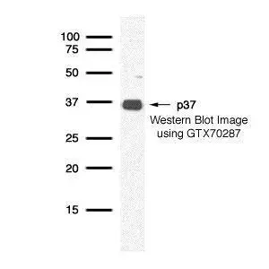
![Various whole cell extracts (30 μg) were separated by 10% SDS-PAGE, and the membrane was blotted with p37 antibody [1320] (GTX70285) diluted at 1:500. The HRP-conjugated anti-mouse IgG antibody (GTX213111-01) was used to detect the primary antibody.](https://www.genetex.com/upload/website/prouct_img/normal/GTX70285/GTX70285_40004_20180518_WB_w_23061221_759.webp)
Product group Antibodies
p37 antibody [1320]GTX70285
ApplicationsImmunoFluorescence, Western Blot, ImmunoCytoChemistry
ReactivityHuman, Mouse
TargetRFC4
- SizePrice
Product group Antibodies
p37 antibodyGTX70287
ApplicationsImmunoFluorescence, Western Blot, ImmunoCytoChemistry
ReactivityHuman
TargetRFC4
- SizePrice
![Various whole cell extracts (30 μg) were separated by 10% SDS-PAGE, and the membrane was blotted with RFC4 antibody [N1C3] (GTX104052) diluted at 1:500. The HRP-conjugated anti-rabbit IgG antibody (GTX213110-01) was used to detect the primary antibody.](https://www.genetex.com/upload/website/prouct_img/normal/GTX104052/GTX104052_40079_20180518_WB_w_23060120_532.webp)
Product group Antibodies
RFC4 antibody [N1C3]GTX104052
ApplicationsImmunoFluorescence, Western Blot, ImmunoCytoChemistry, ImmunoHistoChemistry, ImmunoHistoChemistry Paraffin
ReactivityHuman, Mouse
TargetRFC4
- SizePrice