

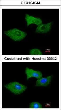
Immunofluorescence analysis of methanol-fixed A549, using RGS2(GTX104944) antibody at 1:200 dilution.
 diluted at 1:500.
Antigen Retrieval: Citrate buffer, pH 6.0, 15 min")
 was separated by 12% SDS-PAGE, and the membrane was blotted with RGS2 antibody (GTX104944) diluted at 1:1000. The HRP-conjugated anti-rabbit IgG antibody (GTX213110-01) was used to detect the primary antibody.")
 was separated by 12% SDS-PAGE, and the membrane was blotted with RGS2 antibody (GTX104944) diluted at 1:1000. The HRP-conjugated anti-rabbit IgG antibody (GTX213110-01) was used to detect the primary antibody.")
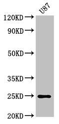
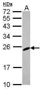
 were separated by 12% SDS-PAGE, and the membrane was blotted with RGS2 antibody (GTX104944) diluted at 1:2500. The HRP-conjugated anti-rabbit IgG antibody (GTX213110-01) was used to detect the primary antibody. (WCE: whole cell extract; ME: membrane extract)")
 were separated by 12% SDS-PAGE, and the membrane was blotted with RGS2 antibody (GTX104944) diluted at 1:1000. The HRP-conjugated anti-rabbit IgG antibody (GTX213110-01) was used to detect the primary antibody.")
Immunofluorescence analysis of methanol-fixed A549, using RGS2(GTX104944) antibody at 1:200 dilution.
RGS2 antibody
GTX104944
ApplicationsImmunoFluorescence, Western Blot, ImmunoCytoChemistry, ImmunoHistoChemistry, ImmunoHistoChemistry Paraffin
Product group Antibodies
ReactivityHuman, Mouse, Rat
TargetRGS2
Overview
- SupplierGeneTex
- Product NameRGS2 antibody
- Delivery Days Customer9
- Application Supplier NoteWB: 1:500-1:3000. ICC/IF: 1:100-1:1000. IHC-P: 1:100-1:1000. *Optimal dilutions/concentrations should be determined by the researcher.Not tested in other applications.
- ApplicationsImmunoFluorescence, Western Blot, ImmunoCytoChemistry, ImmunoHistoChemistry, ImmunoHistoChemistry Paraffin
- CertificationResearch Use Only
- ClonalityPolyclonal
- Concentration0.53 mg/ml
- ConjugateUnconjugated
- Gene ID5997
- Target nameRGS2
- Target descriptionregulator of G protein signaling 2
- Target synonymsG0S8, regulator of G-protein signaling 2, G0 to G1 switch regulatory 8, 24kD, G0/G1 switch regulatory protein 8, cell growth-inhibiting gene 31 protein, cell growth-inhibiting protein 31, regulator of G-protein signaling 2, 24kDa
- HostRabbit
- IsotypeIgG
- Protein IDP41220
- Protein NameRegulator of G-protein signaling 2
- Scientific DescriptionRegulator of G protein signaling (RGS) family members are regulatory molecules that act as GTPase activating proteins (GAPs) for G alpha subunits of heterotrimeric G proteins. RGS proteins are able to deactivate G protein subunits of the Gi alpha, Go alpha and Gq alpha subtypes. They drive G proteins into their inactive GDP-bound forms. Regulator of G protein signaling 2 belongs to this family. The protein acts as a mediator of myeloid differentiation and may play a role in leukemogenesis. [provided by RefSeq]
- ReactivityHuman, Mouse, Rat
- Storage Instruction-20°C or -80°C,2°C to 8°C
- UNSPSC41116161
Datasheet
Related products
Product group Antibodies
Anti-RGS2 AntibodyA13660
ApplicationsWestern Blot, ImmunoHistoChemistry
ReactivityHuman
- SizePrice
Product group Antibodies

RGS2 AntibodyLS-C830279
ApplicationsWestern Blot, ELISA, ImmunoHistoChemistry
ReactivityHuman, Mouse, Rat
TargetRGS2
- SizePrice
Product group Antibodies
RGS2 Polyclonal AntibodyBS-0492R
ApplicationsImmunoFluorescence, ELISA, ImmunoCytoChemistry, ImmunoHistoChemistry, ImmunoHistoChemistry Frozen, ImmunoHistoChemistry Paraffin
ReactivityBovine, Chicken, Equine, Human, Mouse, Porcine, Rat, Sheep
TargetRGS2
- SizePrice
Product group Antibodies
RGS2 Polyclonal AntibodyCAC13905
ApplicationsImmunoFluorescence, Western Blot, ELISA, ImmunoHistoChemistry
TargetRGS2
- SizePrice
Product group Antibodies
RGS2 AntibodyCSB-PA019651LA01HU
ApplicationsImmunoFluorescence, Western Blot, ELISA, ImmunoHistoChemistry
ReactivityHuman
TargetRGS2
- SizePrice
Product group Antibodies
RGS2 antibody [N1C3-3]GTX113349
ApplicationsImmunoPrecipitation, Western Blot
ReactivityHuman, Mouse
TargetRGS2
- SizePrice
![RGS2 antibody [HL2987] detects RGS2 protein by immunohistochemical analysis. Sample: Paraffin-embedded mouse ovary. RGS2 stained by RGS2 antibody [HL2987] (GTX640397) diluted at 1:100. Antigen Retrieval: Citrate buffer, pH 6.0, 15 min](https://www.genetex.com/upload/website/prouct_img/normal/GTX640397/GTX640397_T-45418_20240718_IHC-P_M_24072519_746.webp)
Product group Antibodies
RGS2 antibody [HL2987]GTX640397
ApplicationsWestern Blot, ImmunoHistoChemistry, ImmunoHistoChemistry Paraffin
ReactivityHuman, Mouse, Rat
TargetRGS2
- SizePrice
![Whole tissue extract (50 μg) was separated by 12% SDS-PAGE, and the membrane was blotted with RGS2 antibody [HL2988] (GTX640398) diluted at 1:1000. The HRP-conjugated anti-rabbit IgG antibody (GTX213110-01) was used to detect the primary antibody, and the signal was developed with Trident ECL plus-Enhanced.](https://www.genetex.com/upload/website/prouct_img/normal/GTX640398/GTX640398_T-45418_20240531_WB_M_bladder_24060619_958.webp)
Product group Antibodies
RGS2 antibody [HL2988]GTX640398
ApplicationsWestern Blot, ImmunoHistoChemistry, ImmunoHistoChemistry Paraffin
ReactivityHuman, Mouse
TargetRGS2
- SizePrice

Product group Antibodies
Anti-RGS2 AntibodyHPA013385
ApplicationsImmunoCytoChemistry
ReactivityHuman
TargetRGS2
- SizePrice
Product group Antibodies
Anti-NIPSNAP1 AntibodyCAB17951
ApplicationsWestern Blot, ELISA
ReactivityHuman
TargetRGS2
- SizePrice