

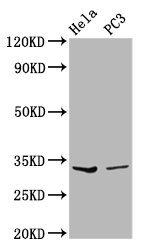
Western Blot Positive WB detected in: Hela whole cell lysate, PC-3 whole cell lysate All lanes: RNASEH1 antibody at 3.59microg/ml Secondary Goat polyclonal to rabbit IgG at 1/50000 dilution Predicted band size: 33 kDa Observed band size: 33 kDa
. Section was blocked with 10% normal goat serum 30min at RT. Then primary antibody (1% BSA) was incubated at 4°C overnight. The primary is detected by a biotinylated secondary antibody and visualized using an HRP conjugated SP system.")
. Section was blocked with 10% normal goat serum 30min at RT. Then primary antibody (1% BSA) was incubated at 4°C overnight. The primary is detected by a biotinylated secondary antibody and visualized using an HRP conjugated SP system.")
Western Blot Positive WB detected in: Hela whole cell lysate, PC-3 whole cell lysate All lanes: RNASEH1 antibody at 3.59microg/ml Secondary Goat polyclonal to rabbit IgG at 1/50000 dilution Predicted band size: 33 kDa Observed band size: 33 kDa
RNASEH1 Antibody
CSB-PA019801LA01HU
ApplicationsWestern Blot, ELISA, ImmunoHistoChemistry
Product group Antibodies
ReactivityHuman
TargetRNASEH1
Overview
- SupplierCusabio
- Product NameRNASEH1 Antibody
- Delivery Days Customer20
- ApplicationsWestern Blot, ELISA, ImmunoHistoChemistry
- CertificationResearch Use Only
- ClonalityPolyclonal
- ConjugateUnconjugated
- Gene ID246243
- Target nameRNASEH1
- Target descriptionribonuclease H1
- Target synonymsH1RNA, PEOB2, RNH1, ribonuclease H1, ribonuclease H type II
- HostRabbit
- IsotypeIgG
- Protein IDO60930
- Protein NameRibonuclease H1
- Scientific DescriptionEndonuclease that specifically degrades the RNA of RNA-DNA hybrids (PubMed:10497183). Plays a role in RNA polymerase II (RNAp II) transcription termination by degrading R-loop RNA-DNA hybrid formation at G-rich pause sites located downstream of the poly(A) site and behind the elongating RNAp II (PubMed:21700224).
- ReactivityHuman
- Storage Instruction-20°C or -80°C
- UNSPSC41116161
Related products
Product group Antibodies
Rnaseh1 Polyclonal AntibodyCAC11443
ApplicationsWestern Blot, ELISA, ImmunoHistoChemistry
TargetRNASEH1
- SizePrice

Product group Antibodies
Anti-RNASEH1 Antibody Picoband(r)A06342-1-CARRIER-FREE
ApplicationsFlow Cytometry, ImmunoFluorescence, Western Blot, ELISA, ImmunoCytoChemistry
ReactivityHuman, Mouse
TargetRNASEH1
- SizePrice

Product group Antibodies
Anti-RNASEH1 AntibodyHPA034817
ApplicationsWestern Blot, ImmunoCytoChemistry
ReactivityHuman
TargetRNASEH1
- SizePrice
Product group Antibodies

RNASEH1 AntibodyLS-C410650
ApplicationsImmunoPrecipitation, Western Blot
ReactivityHuman, Mouse, Rat
TargetRNASEH1
- SizePrice
![Various whole cell extracts (30 μg) were separated by 12% SDS-PAGE, and the membrane was blotted with RNase H1 antibody [N2C3] (GTX117624) diluted at 1:1000. The HRP-conjugated anti-rabbit IgG antibody (GTX213110-01) was used to detect the primary antibody.](https://www.genetex.com/upload/website/prouct_img/normal/GTX117624/GTX117624_44980_20230317_WB_23032819_551.webp)
Product group Antibodies
RNase H1 antibody [N2C3]GTX117624
ApplicationsWestern Blot, ImmunoHistoChemistry, ImmunoHistoChemistry Paraffin
ReactivityHuman, Zebra Fish
TargetRNASEH1
- SizePrice