

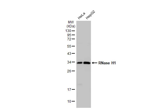
Various whole cell extracts (30 μg) were separated by 12% SDS-PAGE, and the membrane was blotted with RNase H1 antibody [N2C3] (GTX117624) diluted at 1:1000. The HRP-conjugated anti-rabbit IgG antibody (GTX213110-01) was used to detect the primary antibody.
![Whole zebrafish extract (30 μg) was separated by 12% SDS-PAGE, and the membrane was blotted with RNase H1 antibody [N2C3] (GTX117624) diluted at 1:1000. The HRP-conjugated anti-rabbit IgG antibody (GTX213110-01) was used to detect the primary antibody.](https://www.genetex.com/upload/website/prouct_img/normal/GTX117624/GTX117624_40401_20160428_WB_Z_w_23060519_810.webp "Whole zebrafish extract (30 μg) was separated by 12% SDS-PAGE, and the membrane was blotted with RNase H1 antibody [N2C3] (GTX117624) diluted at 1:1000. The HRP-conjugated anti-rabbit IgG antibody (GTX213110-01) was used to detect the primary antibody.")
 antibody at 1:500 dilution.
Antigen Retrieval: Trilogy? (EDTA based, pH 8.0) buffer, 15min")


Various whole cell extracts (30 μg) were separated by 12% SDS-PAGE, and the membrane was blotted with RNase H1 antibody [N2C3] (GTX117624) diluted at 1:1000. The HRP-conjugated anti-rabbit IgG antibody (GTX213110-01) was used to detect the primary antibody.
RNase H1 antibody [N2C3]
GTX117624
ApplicationsWestern Blot, ImmunoHistoChemistry, ImmunoHistoChemistry Paraffin
Product group Antibodies
ReactivityHuman, Zebra Fish
TargetRNASEH1
Overview
- SupplierGeneTex
- Product NameRNase H1 antibody [N2C3]
- Delivery Days Customer9
- Application Supplier NoteWB: 1:500-1:3000. IHC-P: 1:100-1:1000. *Optimal dilutions/concentrations should be determined by the researcher.Not tested in other applications.
- ApplicationsWestern Blot, ImmunoHistoChemistry, ImmunoHistoChemistry Paraffin
- CertificationResearch Use Only
- ClonalityPolyclonal
- Concentration1.3 mg/ml
- ConjugateUnconjugated
- Gene ID246243
- Target nameRNASEH1
- Target descriptionribonuclease H1
- Target synonymsH1RNA, PEOB2, RNH1, ribonuclease H1, ribonuclease H type II
- HostRabbit
- IsotypeIgG
- Protein IDO60930
- Protein NameRibonuclease H1
- ReactivityHuman, Zebra Fish
- Storage Instruction-20°C or -80°C,2°C to 8°C
- UNSPSC41116161
Datasheet
Related products
Product group Antibodies
RNASEH1 AntibodyCSB-PA019801LA01HU
ApplicationsWestern Blot, ELISA, ImmunoHistoChemistry
ReactivityHuman
TargetRNASEH1
- SizePrice
Product group Antibodies
Rnaseh1 Polyclonal AntibodyCAC11443
ApplicationsWestern Blot, ELISA, ImmunoHistoChemistry
TargetRNASEH1
- SizePrice

Product group Antibodies
Anti-RNASEH1 Antibody Picoband(r)A06342-1-CARRIER-FREE
ApplicationsFlow Cytometry, ImmunoFluorescence, Western Blot, ELISA, ImmunoCytoChemistry
ReactivityHuman, Mouse
TargetRNASEH1
- SizePrice

Product group Antibodies
Anti-RNASEH1 AntibodyHPA034817
ApplicationsWestern Blot, ImmunoCytoChemistry
ReactivityHuman
TargetRNASEH1
- SizePrice
Product group Antibodies

RNASEH1 AntibodyLS-C410650
ApplicationsImmunoPrecipitation, Western Blot
ReactivityHuman, Mouse, Rat
TargetRNASEH1
- SizePrice
![Mouse tissue extract (50 μg) was separated by 12% SDS-PAGE, and the membrane was blotted with RNase H1 antibody [HL2343] (GTX638546) diluted at 1:2000. The HRP-conjugated anti-rabbit IgG antibody (GTX213110-01) was used to detect the primary antibody.](https://www.genetex.com/upload/website/prouct_img/normal/GTX638546/GTX638546_T-45026_20230428_WB_M_cerebellum_23050223_492.webp)
Product group Antibodies
RNase H1 antibody [HL2343]GTX638546
ApplicationsWestern Blot, ImmunoHistoChemistry, ImmunoHistoChemistry Paraffin
ReactivityCanine, Human, Mouse
TargetRNASEH1
- SizePrice