

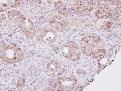
Immunohistochemical analysis of paraffin-embedded NCIN87 xenograft , using RPL8(GTX101614) antibody at 1:100 dilution.
Antigen Retrieval: Citrate buffer, pH 6.0, 15 min
 antibody at 1:200 dilution.")
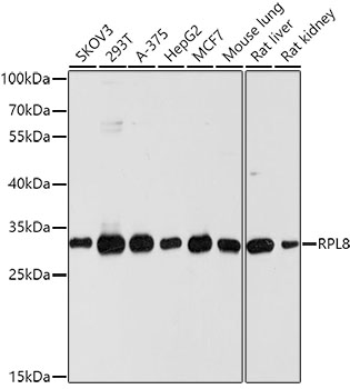
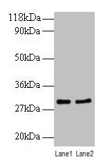
 A: HepG2 (GTX27900) B: Molt-4 (GTX27912) 12% SDS PAGE GTX101614 diluted at 1:1000 The HRP-conjugated anti-rabbit IgG antibody (GTX213110-01) was used to detect the primary antibody.")
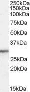
 A: Mouse brain 12% SDS PAGE GTX101614 diluted at 1:1000 The HRP-conjugated anti-rabbit IgG antibody (GTX213110-01) was used to detect the primary antibody.")
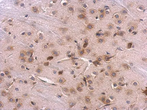
Immunohistochemical analysis of paraffin-embedded NCIN87 xenograft , using RPL8(GTX101614) antibody at 1:100 dilution.
Antigen Retrieval: Citrate buffer, pH 6.0, 15 min
RPL8 antibody
GTX101614
ApplicationsImmunoFluorescence, Western Blot, ImmunoCytoChemistry, ImmunoHistoChemistry, ImmunoHistoChemistry Paraffin
Product group Antibodies
ReactivityHuman, Mouse
TargetRPL8
Overview
- SupplierGeneTex
- Product NameRPL8 antibody
- Delivery Days Customer9
- Application Supplier NoteWB: 1:500-1:3000. ICC/IF: 1:100-1:1000. IHC-P: 1:100-1:1000. *Optimal dilutions/concentrations should be determined by the researcher.Not tested in other applications.
- ApplicationsImmunoFluorescence, Western Blot, ImmunoCytoChemistry, ImmunoHistoChemistry, ImmunoHistoChemistry Paraffin
- CertificationResearch Use Only
- ClonalityPolyclonal
- Concentration0.27 mg/ml
- ConjugateUnconjugated
- Gene ID6132
- Target nameRPL8
- Target descriptionribosomal protein L8
- Target synonymsL8, uL2, large ribosomal subunit protein uL2, 60S ribosomal protein L8
- HostRabbit
- IsotypeIgG
- Protein IDP62917
- Protein NameLarge ribosomal subunit protein uL2
- Scientific DescriptionRibosomes, the organelles that catalyze protein synthesis, consist of a small 40S subunit and a large 60S subunit. Together these subunits are composed of 4 RNA species and approximately 80 structurally distinct proteins. This gene encodes a ribosomal protein that is a component of the 60S subunit. The protein belongs to the L2P family of ribosomal proteins. It is located in the cytoplasm. In rat, the protein associates with the 5.8S rRNA, very likely participates in the binding of aminoacyl-tRNA, and is a constituent of the elongation factor 2-binding site at the ribosomal subunit interface. Alternatively spliced transcript variants encoding the same protein exist. As is typical for genes encoding ribosomal proteins, there are multiple processed pseudogenes of this gene dispersed through the genome. [provided by RefSeq]
- ReactivityHuman, Mouse
- Storage Instruction-20°C or -80°C,2°C to 8°C
- UNSPSC41116161
Datasheet
Related products

Product group Antibodies
Anti-RPL8 Antibody Picoband(r)A06793-1-CARRIER-FREE
ApplicationsWestern Blot, ELISA, ImmunoHistoChemistry
ReactivityHuman, Rat
TargetRPL8
- SizePrice
Product group Antibodies
Anti-RPL8 AntibodyA12769
ApplicationsImmunoFluorescence, Western Blot, ImmunoCytoChemistry, ImmunoHistoChemistry
ReactivityHuman, Mouse, Rat
- SizePrice
Product group Antibodies
Anti-RPL8 Antibody144-10042
ApplicationsImmunoFluorescence, Western Blot
ReactivityHuman, Mouse, Rat
TargetRPL8
- SizePrice
Product group Antibodies

ApplicationsWestern Blot, ELISA, ImmunoHistoChemistry
ReactivityCanine, Human, Mouse, Rat
TargetRPL8
- SizePrice
Product group Antibodies
Rpl8 Polyclonal AntibodyCAC08333
ApplicationsWestern Blot, ELISA
TargetRPL8
- SizePrice
Product group Antibodies
RPL8 AntibodyCSB-PA00595A0RB
ApplicationsWestern Blot, ELISA
ReactivityHuman
TargetRPL8
- SizePrice
Product group Antibodies

RPL8 / Ribosomal Protein L8 AntibodyLS-C496753
ApplicationsWestern Blot
ReactivityHuman, Mouse, Rat
TargetRPL8
- SizePrice
Product group Antibodies
RPL8 antibody, InternalGTX88990
ApplicationsWestern Blot, ImmunoHistoChemistry, ImmunoHistoChemistry Paraffin
ReactivityHuman
TargetRPL8
- SizePrice

Product group Antibodies
Anti-RPL8 AntibodyHPA050165
ApplicationsWestern Blot, ImmunoCytoChemistry, ImmunoHistoChemistry
ReactivityHuman
TargetRPL8
- SizePrice
Product group Antibodies
RPL8 antibodyGTX117934
ApplicationsImmunoFluorescence, Western Blot, ImmunoCytoChemistry, ImmunoHistoChemistry, ImmunoHistoChemistry Paraffin
ReactivityHuman, Mouse, Rat
TargetRPL8
- SizePrice