

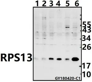
WB analysis of various samples using GTX66837 RPS13 antibody. Lane1 : CT26 whole cell lysate(40ug)
Lane2 : PC12 whole cell lysate(40ug)
Lane3 : MCF-7 whole cell lysate(40ug)
Lane4 : A549 whole cell lysate(40ug)
Lane5 : HEK293T whole cell lysate(40ug)
Lane6 : SGC7901 whole cell lysate(40ug) Dilution : 1:500
RPS13 antibody
GTX66837
ApplicationsWestern Blot
Product group Antibodies
ReactivityHuman, Mouse, Rat
TargetRPS13
Overview
- SupplierGeneTex
- Product NameRPS13 antibody
- Delivery Days Customer9
- Application Supplier NoteWB: 1:500-1:1000. *Optimal dilutions/concentrations should be determined by the researcher.Not tested in other applications.
- ApplicationsWestern Blot
- CertificationResearch Use Only
- ClonalityPolyclonal
- Concentration1 mg/ml
- ConjugateUnconjugated
- Gene ID6207
- Target nameRPS13
- Target descriptionribosomal protein S13
- Target synonymsS13, uS15, small ribosomal subunit protein uS15, 40S ribosomal protein S13
- HostRabbit
- IsotypeIgG
- Protein IDP62277
- Protein NameSmall ribosomal subunit protein uS15
- Scientific DescriptionRibosomes, the organelles that catalyze protein synthesis, consist of a small 40S subunit and a large 60S subunit. Together these subunits are composed of 4 RNA species and approximately 80 structurally distinct proteins. This gene encodes a ribosomal protein that is a component of the 40S subunit. The protein belongs to the S15P family of ribosomal proteins. It is located in the cytoplasm. The protein has been shown to bind to the 5.8S rRNA in rat. The gene product of the E. coli ortholog (ribosomal protein S15) functions at early steps in ribosome assembly. This gene is co-transcribed with two U14 small nucleolar RNA genes, which are located in its third and fifth introns. As is typical for genes encoding ribosomal proteins, there are multiple processed pseudogenes of this gene dispersed through the genome. [provided by RefSeq, Jul 2008]
- ReactivityHuman, Mouse, Rat
- Storage Instruction-20°C or -80°C,2°C to 8°C
- UNSPSC41116161
Datasheet
Related products
Product group Antibodies
RPS13 AntibodyCSB-PA02485A0RB
ApplicationsWestern Blot, ELISA, ImmunoHistoChemistry
ReactivityHuman
TargetRPS13
- SizePrice

Product group Antibodies
Anti-RPS13 AntibodyA96997
ApplicationsELISA, ImmunoHistoChemistry
ReactivityHuman, Mouse, Rat
- SizePrice

Product group Antibodies
Anti-RPS13 Antibody Picoband(r)A06221-3-CARRIER-FREE
ApplicationsFlow Cytometry, Western Blot, ELISA, ImmunoHistoChemistry
ReactivityHuman
TargetRPS13
- SizePrice
Product group Antibodies
Anti-RPS13 AntibodyHPA005985
ApplicationsImmunoCytoChemistry, ImmunoHistoChemistry
ReactivityHuman
TargetRPS13
- SizePrice
Product group Antibodies

ApplicationsWestern Blot, ImmunoHistoChemistry, ImmunoHistoChemistry Paraffin
ReactivityBovine, Chicken, Human, Monkey, Mouse, Rat, Zebra Fish
TargetRPS13
- SizePrice
Product group Antibodies
RPS13 Polyclonal AntibodyCAC13981
ApplicationsWestern Blot, ELISA, ImmunoHistoChemistry
TargetRPS13
- SizePrice
Product group Antibodies
Anti-RPS13 Antibody144-61165
ApplicationsWestern Blot
ReactivityHuman, Mouse, Rat
TargetRPS13
- SizePrice