

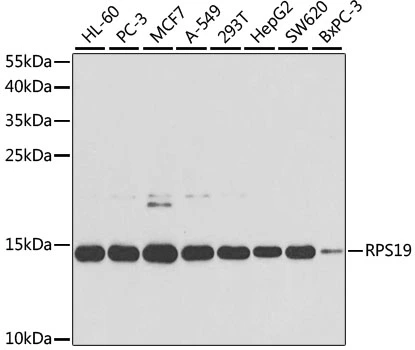
WB analysis of various sample lysates using GTX54725 RPS19 antibody. Dilution : 1:1000 Loading : 25μg per lane
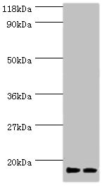
WB analysis of various sample lysates using GTX54725 RPS19 antibody. Dilution : 1:1000 Loading : 25μg per lane
RPS19 antibody
GTX54725
ApplicationsWestern Blot
Product group Antibodies
ReactivityHuman
TargetRPS19
Overview
- SupplierGeneTex
- Product NameRPS19 antibody
- Delivery Days Customer7
- Application Supplier NoteWB: 1:500 - 1:2000. *Optimal dilutions/concentrations should be determined by the researcher.Not tested in other applications.
- ApplicationsWestern Blot
- CertificationResearch Use Only
- ClonalityPolyclonal
- ConjugateUnconjugated
- Gene ID6223
- Target nameRPS19
- Target descriptionribosomal protein S19
- Target synonymsDBA, DBA1, LOH19CR1, S19, eS19, small ribosomal subunit protein eS19, 40S ribosomal protein S19, loss of heterozygosity on chromosome 19, region 1, loss of heterozygosity, 19, chromosomal region 1
- HostRabbit
- IsotypeIgG
- Protein IDP39019
- Protein NameSmall ribosomal subunit protein eS19
- Scientific DescriptionRibosomes, the organelles that catalyze protein synthesis, consist of a small 40S subunit and a large 60S subunit. Together these subunits are composed of 4 RNA species and approximately 80 structurally distinct proteins. This gene encodes a ribosomal protein that is a component of the 40S subunit. The protein belongs to the S19E family of ribosomal proteins. It is located in the cytoplasm. Mutations in this gene cause Diamond-Blackfan anemia (DBA), a constitutional erythroblastopenia characterized by absent or decreased erythroid precursors, in a subset of patients. This suggests a possible extra-ribosomal function for this gene in erythropoietic differentiation and proliferation, in addition to its ribosomal function. Higher expression levels of this gene in some primary colon carcinomas compared to matched normal colon tissues has been observed. As is typical for genes encoding ribosomal proteins, there are multiple processed pseudogenes of this gene dispersed through the genome. [provided by RefSeq, Jul 2008]
- ReactivityHuman
- Storage Instruction-20°C or -80°C,2°C to 8°C
- UNSPSC41116161
Datasheet
Related products
Product group Antibodies
Anti-RPS19 AntibodyA95793
ApplicationsWestern Blot, ELISA, ImmunoHistoChemistry
ReactivityHuman, Mouse, Rat
- SizePrice
Product group Antibodies
Anti-RPS19 Antibody144-02019
ApplicationsWestern Blot
ReactivityHuman, Mouse
TargetRPS19
- SizePrice

Product group Antibodies
Anti-RPS19 Antibody Picoband(r)A02343-1-CARRIER-FREE
ApplicationsFlow Cytometry, ImmunoFluorescence, Western Blot, ELISA, ImmunoCytoChemistry
ReactivityHuman, Mouse, Rat
TargetRPS19
- SizePrice

Product group Antibodies
RPS19 Recombinant AntibodyBSM-54242R
ApplicationsImmunoFluorescence, ImmunoPrecipitation, Western Blot, ImmunoHistoChemistry, ImmunoHistoChemistry Frozen, ImmunoHistoChemistry Paraffin
ReactivityHuman, Mouse, Rat
TargetRPS19
- SizePrice
Product group Antibodies
RPS19 AntibodyCSB-PA00655A0RB
ApplicationsWestern Blot, ELISA, ImmunoHistoChemistry
ReactivityHuman
TargetRPS19
- SizePrice
Product group Antibodies

Goat anti-RPS19EB07023
ApplicationsWestern Blot, ELISA, ImmunoHistoChemistry
ReactivityBovine, Canine, Human, Mouse, Rat
TargetRPS19
- SizePrice
Product group Antibodies
Rps19 Polyclonal AntibodyCAC08354
ApplicationsWestern Blot, ELISA, ImmunoHistoChemistry
TargetRPS19
- SizePrice
Product group Antibodies

RPS19 / Ribosomal Protein S19 AntibodyLS-C331827
ApplicationsWestern Blot, ImmunoHistoChemistry
ReactivityHuman, Mouse
TargetRPS19
- SizePrice
Product group Antibodies
RPS19 antibody, InternalGTX89461
ApplicationsWestern Blot
ReactivityHuman
TargetRPS19
- SizePrice

Product group Antibodies
Anti-RPS19 AntibodyHPA063217
ApplicationsImmunoCytoChemistry
ReactivityHuman
TargetRPS19
- SizePrice