

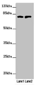
Western blot All lanes: RUFY2 antibody at 1.69ug/ml Lane 1: PC-3 whole cell lysate Lane 2: Mouse spleen tissue Secondary Goat polyclonal to rabbit IgG at 1/10000 dilution Predicted band size: 76, 71, 75, 47, 44 kDa Observed band size: 76 kDa


Western blot All lanes: RUFY2 antibody at 1.69ug/ml Lane 1: PC-3 whole cell lysate Lane 2: Mouse spleen tissue Secondary Goat polyclonal to rabbit IgG at 1/10000 dilution Predicted band size: 76, 71, 75, 47, 44 kDa Observed band size: 76 kDa
RUFY2 Antibody
CSB-PA848835ESR1HU
ApplicationsWestern Blot, ELISA, ImmunoHistoChemistry
Product group Antibodies
ReactivityHuman, Mouse
TargetRUFY2
Overview
- SupplierCusabio
- Product NameRUFY2 Antibody
- Delivery Days Customer20
- ApplicationsWestern Blot, ELISA, ImmunoHistoChemistry
- CertificationResearch Use Only
- ClonalityPolyclonal
- ConjugateUnconjugated
- Gene ID55680
- Target nameRUFY2
- Target descriptionRUN and FYVE domain containing 2
- Target synonymsRABIP4R, ZFYVE13, RUN and FYVE domain-containing protein 2, Run- and FYVE-domain containing protein, antigen MU-RMS-40.17, rab4-interacting protein related
- HostRabbit
- IsotypeIgG
- Protein IDQ8WXA3
- Protein NameRUN and FYVE domain-containing protein 2
- ReactivityHuman, Mouse
- Storage Instruction-20°C or -80°C
- UNSPSC41116161
Related products

Product group Antibodies
Anti-RUFY2 Antibody Picoband(r)A13178-1-CARRIER-FREE
ApplicationsFlow Cytometry, ImmunoFluorescence, Western Blot, ELISA, ImmunoCytoChemistry
ReactivityHuman
TargetRUFY2
- SizePrice
Product group Antibodies

RUFY2 AntibodyLS-C409527
ApplicationsWestern Blot, ImmunoHistoChemistry
ReactivityHuman, Mouse, Rat
TargetRUFY2
- SizePrice

Product group Antibodies
Anti-RUFY2 AntibodyHPA039792
ApplicationsWestern Blot, ImmunoCytoChemistry, ImmunoHistoChemistry
ReactivityHuman
TargetRUFY2
- SizePrice