

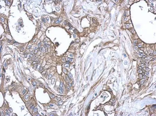
Immunohistochemical analysis of paraffin-embedded human gastric cancer, using S100A10(GTX100697) antibody at 1:500 dilution.
Antigen Retrieval: Trilogy? (EDTA based, pH 8.0) buffer, 15min
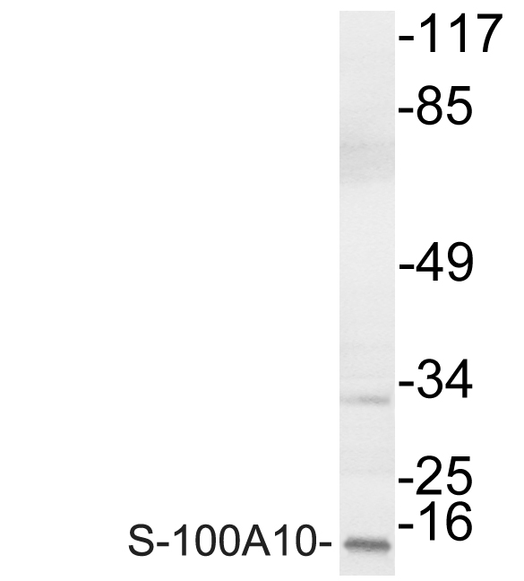
 A: A431 (GTX27909) 15% SDS PAGE GTX100697 diluted at 1:1000")
 and S100A10 knockout (KO) 293T cell extracts (30 μg) were separated by 15% SDS-PAGE, and the membrane was blotted with S100A10 antibody (GTX100697) diluted at 1:1000. The HRP-conjugated anti-rabbit IgG antibody (GTX213110-01) was used to detect the primary antibody, and the signal was developed with Trident ECL plus-Enhanced.")
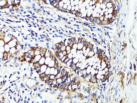
Immunohistochemical analysis of paraffin-embedded human gastric cancer, using S100A10(GTX100697) antibody at 1:500 dilution.
Antigen Retrieval: Trilogy? (EDTA based, pH 8.0) buffer, 15min
S100A10 antibody
GTX100697
ApplicationsWestern Blot, ImmunoHistoChemistry, ImmunoHistoChemistry Frozen, ImmunoHistoChemistry Paraffin
Product group Antibodies
ReactivityHuman, Mouse
TargetS100A10
Overview
- SupplierGeneTex
- Product NameS100A10 antibody
- Delivery Days Customer9
- Application Supplier NoteWB: 1:500-1:3000. IHC-P: 1:100-1:1000. *Optimal dilutions/concentrations should be determined by the researcher.Not tested in other applications.
- ApplicationsWestern Blot, ImmunoHistoChemistry, ImmunoHistoChemistry Frozen, ImmunoHistoChemistry Paraffin
- CertificationResearch Use Only
- ClonalityPolyclonal
- Concentration1 mg/ml
- ConjugateUnconjugated
- Gene ID6281
- Target nameS100A10
- Target descriptionS100 calcium binding protein A10
- Target synonyms42C, ANX2L, ANX2LG, CAL1L, CLP11, Ca[1], GP11, P11, p10, protein S100-A10, S100 calcium binding protein A10 (annexin II ligand, calpactin I, light polypeptide (p11)), annexin II ligand, calpactin I, light polypeptide, annexin II tetramer (AIIt) p11 subunit, calpactin I light chain, calpactin-1 light chain, cellular ligand of annexin II
- HostRabbit
- IsotypeIgG
- Protein IDP60903
- Protein NameProtein S100-A10
- Scientific DescriptionThe protein encoded by this gene is a member of the S100 family of proteins containing 2 EF-hand calcium-binding motifs. S100 proteins are localized in the cytoplasm and/or nucleus of a wide range of cells, and involved in the regulation of a number of cellular processes such as cell cycle progression and differentiation. S100 genes include at least 13 members which are located as a cluster on chromosome 1q21. This protein may function in exocytosis and endocytosis. [provided by RefSeq]
- ReactivityHuman, Mouse
- Storage Instruction-20°C or -80°C,2°C to 8°C
- UNSPSC41116161
Datasheet
Related products
Product group Antibodies
Anti-S-100A10 AntibodyA95324
ApplicationsWestern Blot, ELISA, ImmunoHistoChemistry
ReactivityHuman, Mouse, Rat
- SizePrice

Product group Antibodies
Anti-S100A10 Antibody Picoband(r)A02787-2-CARRIER-FREE
ApplicationsFlow Cytometry, ImmunoFluorescence, Western Blot, ELISA, ImmunoCytoChemistry, ImmunoHistoChemistry
ReactivityHuman, Monkey, Mouse, Rat
TargetS100A10
- SizePrice
Product group Antibodies
Anti-S100A10 Antibody144-60742
ApplicationsWestern Blot, ImmunoHistoChemistry
ReactivityHuman, Monkey, Mouse, Rat
TargetS100A10
- SizePrice
Product group Antibodies

S100A10 AntibodyLS-C749651
ApplicationsWestern Blot, ImmunoHistoChemistry
ReactivityHuman, Monkey, Mouse, Rat
TargetS100A10
- SizePrice
Product group Antibodies
S100A10 Recombinant Antibody, AbBy Fluor-350 ConjugatedBSM-61398R-BF350
ApplicationsImmunoFluorescence, Western Blot
ReactivityHuman, Mouse
TargetS100A10
- SizePrice
Product group Antibodies
S100A10 AntibodyCSB-PA004043
ApplicationsWestern Blot, ELISA, ImmunoHistoChemistry
ReactivityHuman, Mouse, Rat
TargetS100A10
- SizePrice
Product group Antibodies
ApplicationsImmunoPrecipitation, Western Blot, ImmunoCytoChemistry, ImmunoHistoChemistry
TargetS100A10
- SizePrice
![ICC/IF analysis of HeLa (left) and L-02 (right) cells using GTX83075 S100A10 antibody [4E7E10]. Green : S100A10 Blue: DRAQ5 fluorescent DNA dye Red: Actin filaments](https://www.genetex.com/upload/website/prouct_img/normal/GTX83075/GTX83075_20170912_ICCIF_w_23061322_662.webp)
Product group Antibodies
S100A10 antibody [4E7E10]GTX83075
ApplicationsImmunoFluorescence, Western Blot, ELISA, ImmunoCytoChemistry, ImmunoHistoChemistry, ImmunoHistoChemistry Paraffin
ReactivityHuman
TargetS100A10
- SizePrice

Product group Antibodies
Anti-S100A10 AntibodyHPA003340
ApplicationsWestern Blot, ImmunoCytoChemistry, ImmunoHistoChemistry
ReactivityHuman
TargetS100A10
- SizePrice
Product group Antibodies
S100A10 antibodyGTX65938
ApplicationsWestern Blot, ImmunoHistoChemistry, ImmunoHistoChemistry Paraffin
ReactivityHuman, Monkey, Mouse
TargetS100A10
- SizePrice