

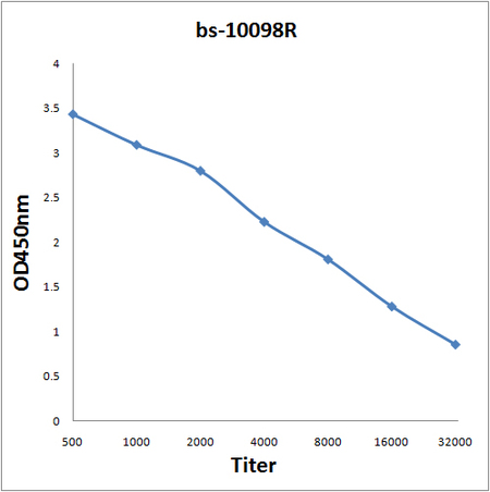
Antigen: bs-10098P, 0.2ug/100ul \nPrimary: Antiserum, 1:500, 1:1000, 1:2000, 1:4000, 1:8000, 1:16000, 1:32000; \nSecondary: HRP conjugated Goat-Anti-Rabbit IgG(bs-0295G-HRP) at 1: 5000;\nTMB staining;\nRead the data in MicroplateReader by 450
Antigen: bs-10098P, 0.2ug/100ul \nPrimary: Antiserum, 1:500, 1:1000, 1:2000, 1:4000, 1:8000, 1:16000, 1:32000; \nSecondary: HRP conjugated Goat-Anti-Rabbit IgG(bs-0295G-HRP) at 1: 5000;\nTMB staining;\nRead the data in MicroplateReader by 450
S100A6 Polyclonal Antibody
BS-10098R
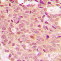
ApplicationsImmunoFluorescence, ELISA, ImmunoCytoChemistry, ImmunoHistoChemistry, ImmunoHistoChemistry Frozen, ImmunoHistoChemistry Paraffin
Product group Antibodies
ReactivityCanine, Equine, Human, Mouse, Porcine, Rabbit, Rat
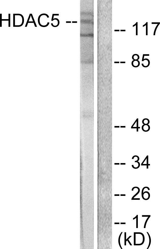
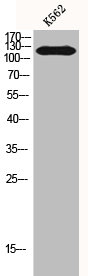
TargetHDAC5
Overview
- SupplierBioss
- Product NameS100A6 Polyclonal Antibody
- Delivery Days Customer16
- ApplicationsImmunoFluorescence, ELISA, ImmunoCytoChemistry, ImmunoHistoChemistry, ImmunoHistoChemistry Frozen, ImmunoHistoChemistry Paraffin
- Applications SupplierELISA(1:500-1000), IHC-P(1:200-400), IHC-F(1:100-500), IF(IHC-P)(1:50-200), IF(IHC-F)(1:50-200), IF(ICC)(1:50-200)
- CertificationResearch Use Only
- ClonalityPolyclonal
- Concentration1 ug/ul
- ConjugateUnconjugated
- Gene ID10014
- Target nameHDAC5
- Target descriptionhistone deacetylase 5
- Target synonymsHD5, NY-CO-9, histone deacetylase 5, antigen NY-CO-9
- HostRabbit
- IsotypeIgG
- ReactivityCanine, Equine, Human, Mouse, Porcine, Rabbit, Rat
- Storage Instruction-20°C
- UNSPSC41116161
Datasheet
Related products
Product group Antibodies
Anti-HDAC5 AntibodyA95225
ApplicationsImmunoFluorescence, Western Blot, ELISA, ImmunoHistoChemistry
ReactivityHuman, Mouse, Rat
- SizePrice

Product group Antibodies
Anti-HDAC5 Antibody Picoband(r)A01230-5-CARRIER-FREE
ApplicationsWestern Blot, ELISA, ImmunoHistoChemistry
ReactivityHuman, Mouse, Rat
TargetHDAC5
- SizePrice
Product group Antibodies
Anti-HDAC5 Antibody144-07189
ApplicationsImmunoFluorescence, Western Blot
ReactivityHuman
TargetHDAC5
- SizePrice

Product group Antibodies
Anti-HDAC5 AntibodyAMAB91501
ApplicationsImmunoCytoChemistry, ImmunoHistoChemistry
ReactivityHuman
TargetHDAC5
- SizePrice
Product group Antibodies

HDAC5 Antibody (Ser498)LS-C769270
ApplicationsImmunoFluorescence, Western Blot, ELISA, ImmunoHistoChemistry, ImmunoHistoChemistry Paraffin
ReactivityHuman, Mouse
TargetHDAC5
- SizePrice
Product group Antibodies
HDAC5 AntibodyCSB-PA002888
ApplicationsImmunoFluorescence, Western Blot, ELISA, ImmunoHistoChemistry
ReactivityHuman, Mouse
TargetHDAC5
- SizePrice
Product group Antibodies
HDAC5 (phospho Ser498) antibodyGTX32287
ApplicationsImmunoFluorescence, Western Blot, ImmunoCytoChemistry, ImmunoHistoChemistry, ImmunoHistoChemistry Paraffin
ReactivityHuman, Mouse, Rat
TargetHDAC5
- SizePrice