

S100A7 / Psoriasin Antibody
LS-C408380
ApplicationsWestern Blot, ImmunoHistoChemistry
Product group Antibodies
ReactivityHuman, Mouse, Rat
TargetS100A7
Overview
- SupplierLifeSpan BioSciences
- Product NameS100A7 / Psoriasin Antibody
- Delivery Days Customer23
- Application Supplier NoteThe predicted MW is 11kDa, while the observed MW by Western blot was Refer to Figures.
- ApplicationsWestern Blot, ImmunoHistoChemistry
- Applications SupplierIHC (1:50 - 1:200), WB (1:500 - 1:2000) The predicted MW is 11kDa, while the observed MW by Western blot was Refer to Figures.
- CertificationResearch Use Only
- ClonalityPolyclonal
- Concentration0.14 mg/ml
- ConjugateUnconjugated
- Estimated Purity...
- Gene ID6278
- Target nameS100A7
- Target descriptionS100 calcium binding protein A7
- Target synonymsPSOR1, S100A7c, protein S100-A7, psoriasin 1
- HostRabbit
- IsotypeIgG
- ReactivityHuman, Mouse, Rat
- Storage Instruction-20°C
- UNSPSC41116161
Related products

Product group Antibodies
Anti-Psoriasin/S100A7 Antibody Picoband(r)A02369-1-CARRIER-FREE
ApplicationsFlow Cytometry, ImmunoFluorescence, Western Blot, ELISA, ImmunoCytoChemistry, ImmunoHistoChemistry
ReactivityHuman, Mouse, Rat
TargetS100A7
- SizePrice
Product group Antibodies
Anti-S100A7 Antibody144-65426
ApplicationsImmunoHistoChemistry
ReactivityHuman, Mouse, Rat
TargetS100A7
- SizePrice
Product group Antibodies

S100A7 / Psoriasin AntibodyLS-C831555
ApplicationsImmunoHistoChemistry
ReactivityHuman
TargetS100A7
- SizePrice
Product group Antibodies
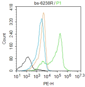
S100A7 Polyclonal AntibodyBS-6238R
ApplicationsFlow Cytometry, ImmunoFluorescence, ELISA, ImmunoCytoChemistry, ImmunoHistoChemistry, ImmunoHistoChemistry Frozen, ImmunoHistoChemistry Paraffin
ReactivityBovine, Equine, Human
TargetS100A7
- SizePrice
Product group Antibodies
S100A7 Polyclonal AntibodyCAC11694
ApplicationsImmunoFluorescence, ELISA, ImmunoHistoChemistry
TargetS100A7
- SizePrice
Product group Antibodies
S100A7 AntibodyCSB-PA020635HA01HU
ApplicationsImmunoFluorescence, ELISA, ImmunoHistoChemistry
ReactivityHuman
TargetS100A7
- SizePrice
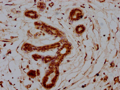
![WB analysis of (A) MCF-10a and (B) MCF-7 cell lysate using GTX13680 Psoriasin antibody [47C1068]. Dilution : 1 μg/ml](https://www.genetex.com/upload/website/prouct_img/normal/GTX13680/GTX13680_975_WB_w_23060620_152.webp)
Product group Antibodies
Psoriasin antibody [47C1068]GTX13680
ApplicationsFlow Cytometry, ImmunoFluorescence, ImmunoPrecipitation, Western Blot, ImmunoCytoChemistry, ImmunoHistoChemistry, ImmunoHistoChemistry Frozen, ImmunoHistoChemistry Paraffin
ReactivityHuman
TargetS100A7
- SizePrice

Product group Antibodies
Anti-S100A7 AntibodyHPA006997
ApplicationsImmunoHistoChemistry
ReactivityHuman
TargetS100A7
- SizePrice
Product group Antibodies
Anti-TOM20 Monoclonal AntibodyCAB19403
ApplicationsImmunoFluorescence, ImmunoPrecipitation, Western Blot, ELISA, ImmunoCytoChemistry, ImmunoHistoChemistry, ImmunoHistoChemistry Paraffin
ReactivityHuman
TargetS100A7
- SizePrice