

S100B / S100 Beta Antibody (C-Terminus, Magnetic beads)
LS-C172094
ApplicationsImmunoPrecipitation
Product group Antibodies
TargetS100B
Overview
- SupplierLifeSpan BioSciences
- Product NameS100B / S100 Beta Antibody (C-Terminus, Magnetic beads)
- Delivery Days Customer23
- ApplicationsImmunoPrecipitation
- Applications SupplierIP
- CertificationResearch Use Only
- ClonalityPolyclonal
- ConjugateBioMagnetic Particle
- Estimated Purity...
- Gene ID6285
- Target nameS100B
- Target descriptionS100 calcium binding protein B
- Target synonymsNEF; protein S100-B; S100; S100 calcium-binding protein, beta (neural); S-100 calcium-binding protein, beta chain; S-100 protein subunit beta; S100-B; S100beta
- HostRabbit
- Storage Instruction2°C to 8°C
- UNSPSC12352203
Related products
Product group Antibodies
References
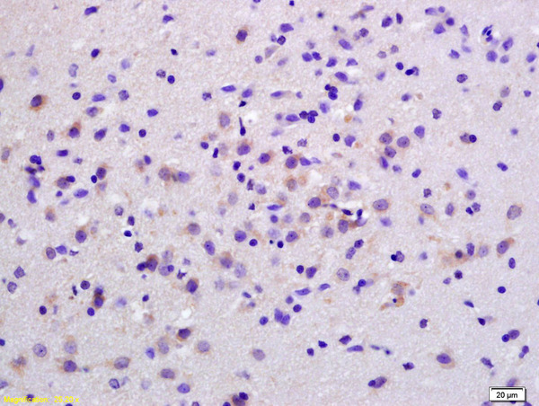
S100B Polyclonal AntibodyBS-2015R
ApplicationsImmunoFluorescence, ELISA, ImmunoCytoChemistry, ImmunoHistoChemistry, ImmunoHistoChemistry Frozen, ImmunoHistoChemistry Paraffin
TargetS100B
- SizePrice

Product group Antibodies
References
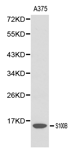
ApplicationsImmunoFluorescence, ImmunoPrecipitation, Western Blot, ImmunoCytoChemistry, ImmunoHistoChemistry
TargetS100B
- SizePrice
Product group Antibodies
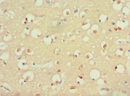
Anti-S100B AntibodyA28225
ApplicationsWestern Blot, ImmunoHistoChemistry
- SizePrice

Product group Antibodies
References
S100 beta antibodyGTX129573
ApplicationsImmunoFluorescence, Western Blot, ImmunoCytoChemistry, ImmunoHistoChemistry, ImmunoHistoChemistry Frozen, ImmunoHistoChemistry Paraffin
TargetS100B
- SizePrice
Product group Antibodies
S100B AntibodyCSB-PA020643ESR1HU
ApplicationsELISA, ImmunoHistoChemistry
ReactivityHuman
TargetS100B
- SizePrice