

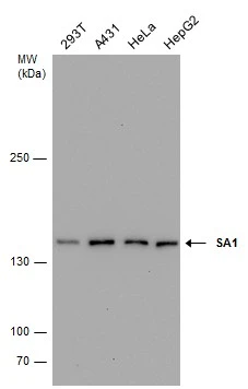
Various whole cell extracts (30 μg) were separated by 5% SDS-PAGE, and the membrane was blotted with SA1 antibody (GTX631989) diluted at 1:1000.
![Non-transfected (–) and transfected (+) 293T whole cell extracts (30 μg) were separated by 5% SDS-PAGE, and the membrane was blotted with SA1 antibody [GT8810] (GTX631989) diluted at 1:500.](https://www.genetex.com/upload/website/prouct_img/normal/GTX631989/GTX631989_41891_20161013_WB_shRNA_watermark_w_23061202_397.webp "Non-transfected (–) and transfected (+) 293T whole cell extracts (30 μg) were separated by 5% SDS-PAGE, and the membrane was blotted with SA1 antibody [GT8810] (GTX631989) diluted at 1:500.")
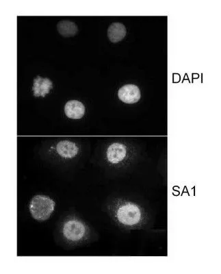
![SA1 antibody [GT8810] detects SA1 protein at nucleus by immunofluorescent analysis. Sample: HeLa cells were fixed in 4% paraformaldehyde at RT for 15 min. Green: SA1 protein stained by SA1 antibody [GT8810] (GTX631989) diluted at 1:1000. Blue: Hoechst 33342 staining. Scale bar = 10 μm.](https://www.genetex.com/upload/website/prouct_img/normal/GTX631989/GTX631989_41891_20141112_IFA_w_23061202_882.webp "SA1 antibody [GT8810] detects SA1 protein at nucleus by immunofluorescent analysis. Sample: HeLa cells were fixed in 4% paraformaldehyde at RT for 15 min. Green: SA1 protein stained by SA1 antibody [GT8810] (GTX631989) diluted at 1:1000. Blue: Hoechst 33342 staining. Scale bar = 10 μm.")
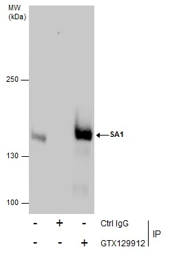
![Immunoprecipitation of SA1 protein from HeLa whole cell extracts using 5 μg of SA1 antibody [GT8810] (GTX631989). Western blot analysis was performed using SA1 antibody [GT8810] (GTX631989) diluted at 1:500. EasyBlot anti-Mouse IgG (GTX221667-01) was used as a secondary reagent.](https://www.genetex.com/upload/website/prouct_img/normal/GTX631989/GTX631989_41891_20160517_IP_w_23061202_721.webp "Immunoprecipitation of SA1 protein from HeLa whole cell extracts using 5 μg of SA1 antibody [GT8810] (GTX631989). Western blot analysis was performed using SA1 antibody [GT8810] (GTX631989) diluted at 1:500. EasyBlot anti-Mouse IgG (GTX221667-01) was used as a secondary reagent.")
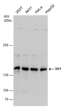
Various whole cell extracts (30 μg) were separated by 5% SDS-PAGE, and the membrane was blotted with SA1 antibody (GTX631989) diluted at 1:1000.
SA1 antibody [GT8810]
GTX631989
ApplicationsImmunoFluorescence, ImmunoPrecipitation, Western Blot, ImmunoCytoChemistry
Product group Antibodies
ReactivityHuman
TargetSTAG1
Overview
- SupplierGeneTex
- Product NameSA1 antibody [GT8810]
- Delivery Days Customer9
- Application Supplier NoteWB: 1:500-1:3000. ICC/IF: 1:100-1:1000. IP: 1:100-1:500. *Optimal dilutions/concentrations should be determined by the researcher.Not tested in other applications.
- ApplicationsImmunoFluorescence, ImmunoPrecipitation, Western Blot, ImmunoCytoChemistry
- CertificationResearch Use Only
- ClonalityMonoclonal
- Clone IDGT8810
- Concentration1 mg/ml
- ConjugateUnconjugated
- Gene ID10274
- Target nameSTAG1
- Target descriptionSTAG1 cohesin complex component
- Target synonymsMRD47, SA1, SCC3A, cohesin subunit SA-1, SCC3 homolog 1, nuclear protein stromal antigen 1, sister chromatid cohesion 3 homolog A, stromal antigen 1
- HostMouse
- IsotypeIgG2a
- Protein IDQ8WVM7
- Protein NameCohesin subunit SA-1
- Scientific DescriptionThis gene is a member of the SCC3 family and is expressed in the nucleus. It encodes a component of cohesin, a multisubunit protein complex that provides sister chromatid cohesion along the length of a chromosome from DNA replication through prophase and prometaphase, after which it is dissociated in preparation for segregation during anaphase. [provided by RefSeq, Jul 2008]
- ReactivityHuman
- Storage Instruction-20°C or -80°C,2°C to 8°C
- UNSPSC41116161
Datasheet
Related products
Product group Antibodies
Anti-STAG1 (Center) Antibody102-22387
ApplicationsWestern Blot
TargetSTAG1
- SizePrice

Product group Antibodies
Anti-SA1/STAG1 Antibody Picoband(r)A06471-1-CARRIER-FREE
ApplicationsFlow Cytometry, ImmunoFluorescence, Western Blot, ELISA, ImmunoCytoChemistry
ReactivityHuman
TargetSTAG1
- SizePrice
Product group Antibodies
ApplicationsImmunoFluorescence, Western Blot, ELISA, ImmunoCytoChemistry, ImmunoHistoChemistry, ImmunoHistoChemistry Frozen, ImmunoHistoChemistry Paraffin
ReactivityBovine, Canine, Chicken, Equine, Human, Mouse, Porcine, Rabbit, Rat
TargetSTAG1
- SizePrice
Product group Antibodies
STAG1 AntibodyCSB-PA022785GA01HU
ApplicationsWestern Blot, ELISA
ReactivityHuman, Mouse, Rat
TargetSTAG1
- SizePrice
Product group Antibodies
SA1 antibodyGTX24455
ApplicationsImmunoFluorescence, Western Blot, ImmunoCytoChemistry, Other Application
ReactivityHuman, Xenopus
TargetSTAG1
- SizePrice
Product group Antibodies

STAG1 / SA1 Antibody (aa1150-1200)LS-C287086
ApplicationsImmunoPrecipitation, Western Blot, ImmunoCytoChemistry
ReactivityHuman
TargetSTAG1
- SizePrice

Product group Antibodies
Anti-STAG1 AntibodyHPA058653
ApplicationsImmunoCytoChemistry
ReactivityHuman
TargetSTAG1
- SizePrice
Product group Antibodies
SA1 antibodyGTX129912
ApplicationsImmunoPrecipitation, Western Blot
ReactivityHuman
TargetSTAG1
- SizePrice
Product group Antibodies
SA1 antibodyGTX130364
ApplicationsImmunoPrecipitation, Western Blot
ReactivityHuman, Mouse, Rat
TargetSTAG1
- SizePrice
![A431 whole cell and nuclear extracts (30 μg) were separated by 5% SDS-PAGE, and the membrane was blotted with SA1 antibody [HL3139] (GTX640631) diluted at 1:1000. The HRP-conjugated anti-rabbit IgG antibody (GTX213110-01) was used to detect the primary antibody.](https://www.genetex.com/upload/website/prouct_img/normal/GTX640631/GTX640631_T-45474_20240802_WB_Fraction_24080602_623.webp)
Product group Antibodies
SA1 antibody [HL3139]GTX640631
ApplicationsWestern Blot
ReactivityHuman
TargetSTAG1
- SizePrice