

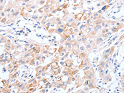
Immunohistochemistry of paraffin-embedded Human thyroid cancer tissue using SCARB1 Polyclonal Antibody at dilution 1:60
Immunohistochemistry of paraffin-embedded Human thyroid cancer tissue using SCARB1 Polyclonal Antibody at dilution 1:60
SCARB1 Polyclonal Antibody
E-AB-10361
ApplicationsImmunoHistoChemistry
Product group Antibodies
TargetSCARB1
Overview
- SupplierElabscience
- Product NameSCARB1 Polyclonal Antibody
- Delivery Days Customer12
- ApplicationsImmunoHistoChemistry
- Applications SupplierELISA IHC
- CertificationResearch Use Only
- ClonalityPolyclonal
- Concentration0.5 mg/ml
- ConjugateUnconjugated
- Gene ID949
- Target nameSCARB1
- Target descriptionscavenger receptor class B member 1
- Target synonymsCD36L1, CLA-1, CLA1, HDLCQ6, HDLQTL6, SR-BI, SRB1, scavenger receptor class B member 1, CD36 and LIMPII analogous 1, CD36 antigen (collagen type I receptor, thrombospondin receptor)-like 1, scavenger receptor class B type III
- HostRabbit
- IsotypeIgG
- Protein IDQ8WTV0
- Protein NameScavenger receptor class B member 1
- Scientific DescriptionThe protein encoded by this gene is a plasma membrane receptor for high density lipoprotein cholesterol (HDL). The encoded protein mediates cholesterol transfer to and from HDL. In addition, this protein is a receptor for hepatitis C virus glycoprotein E2. Two transcript variants encoding different isoforms have been found for this gene.
- Storage Instruction-20°C
- UNSPSC41116161
MSDS
Related products
Product group Antibodies
SCARB1 AntibodyCSB-PA166219
ApplicationsELISA, ImmunoHistoChemistry
ReactivityHuman
TargetSCARB1
- SizePrice

Product group Antibodies
Anti-Scavenging Receptor SR-BI/SCARB1 Antibody Picoband(r)A01093-1-CARRIER-FREE
ApplicationsFlow Cytometry, Western Blot, ELISA
ReactivityHuman, Mouse
TargetSCARB1
- SizePrice
Product group Antibodies

Anti-CD36 [185-1G2 (B467)]Ab01539-1.1
ApplicationsFlow Cytometry, ImmunoFluorescence, ImmunoPrecipitation, ImmunoHistoChemistry, Neutralisation/Blocking
ReactivityHuman
TargetSCARB1
- SizePrice
Product group Antibodies
Anti-SCARB1 AntibodyA29805
ApplicationsWestern Blot, ImmunoHistoChemistry
ReactivityHuman, Mouse, Rat
- SizePrice

Product group Antibodies
Anti-SCARB1 AntibodyHPA066285
ApplicationsImmunoCytoChemistry
ReactivityHuman
TargetSCARB1
- SizePrice
Product group Antibodies

Goat anti-SCARB1 / SR-BIEB12300
ApplicationsWestern Blot, ELISA, ImmunoHistoChemistry
ReactivityHuman
TargetSCARB1
- SizePrice
Product group Antibodies

SCARB1 / SR-BI AntibodyLS-C400786
ApplicationsELISA, ImmunoHistoChemistry
ReactivityHuman
TargetSCARB1
- SizePrice
Product group Antibodies
Scarb1 Polyclonal AntibodyCAC10509
ApplicationsELISA, ImmunoHistoChemistry
TargetSCARB1
- SizePrice