

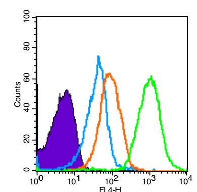
Mouse spleen cells were fixed with 4% PFA (10min at room temperature) and then permeabilized with 90% ice-cold methanol for 20 min. The cells were then incubated in 5% BSA to block non-specific protein-protein interactions for 30 min at room temperature. The cells were then stained with SCF Polyclonal Antibody, Unconjugated (bs-0545R) at 1:33 for 30 min at room temperature. A Goat anti-rabbit IgG-AF647 secondary antibody was used at 1ug for 40 min at room temperature. Acquisition of 10,000 events was performed. Primary antibody staining (green) is compared to compared to unstained cells (purple), secondary only (light blue), and isotype control (orange).
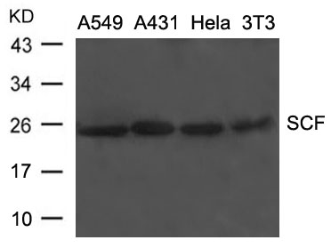
 lysates probed with SCF Polyclonal Antibody, Unconjugated (bs-0545R) at 1:300 dilution and 4˚C overnight incubation. Followed by conjugated secondary antibody incubation at 1:10000 for 60 min at 37˚C.")
 and then were incubated in 5%BSA to block non-specific protein-protein interactions for 30 min at room temperature. The cells were then stained with SCF Polyclonal Antibody, Unconjugated (bs-0545R) at 1:100 for 30 min at room temperature. A Goat anti-rabbit IgG-PE (bs-0295G-PE) secondary antibody was used at 1ug for 40 min at room temperature. Acquisition of 20,000 events was performed. Primary antibody staining (green) is compared to compared to unstained cells (purple), secondary only (light blue), and isotype control (orange).")
 for 15 minutes; Block endogenous peroxidase by 3% hydrogen peroxide for 30 minutes; Blocking buffer (normal goat serum) at 37°C for 20 minutes; Antibody incubation with SCF Polyclonal Antibody, Unconjugated (bs-0545R) at 1:200 overnight at 4°C, followed by a conjugated secondary antibody and DAB staining.")
 for 15 minutes; Block endogenous peroxidase by 3% hydrogen peroxide for 30 minutes; Blocking buffer (normal goat serum) at 37°C for 20 minutes; Antibody incubation with SCF Polyclonal Antibody, Unconjugated (bs-0545R) at 1:200 overnight at 4°C, followed by a conjugated secondary antibody and DAB staining.")
, Unconjugated at 1:200, followed by conjugation to the secondary antibody and DAB staining")
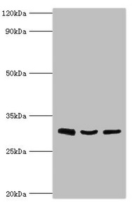
 at 1:1000 dilution and 4˚C overnight incubation. Followed by conjugated secondary antibody incubation at 1:20000 for 60 min at 37˚C.")
 at 37°C for 20 min; Antibody incubation with (SCF) polyclonal Antibody, Unconjugated (bs-0545R) 1:100, 90 minutes at 37°C; followed by a FITC conjugated Goat Anti-Rabbit IgG antibody at 37°C for 90 minutes, DAPI (blue, C02-04002) was used to stain the cell nuclei.")
Mouse spleen cells were fixed with 4% PFA (10min at room temperature) and then permeabilized with 90% ice-cold methanol for 20 min. The cells were then incubated in 5% BSA to block non-specific protein-protein interactions for 30 min at room temperature. The cells were then stained with SCF Polyclonal Antibody, Unconjugated (bs-0545R) at 1:33 for 30 min at room temperature. A Goat anti-rabbit IgG-AF647 secondary antibody was used at 1ug for 40 min at room temperature. Acquisition of 10,000 events was performed. Primary antibody staining (green) is compared to compared to unstained cells (purple), secondary only (light blue), and isotype control (orange).
SCF Polyclonal Antibody
BS-0545R
ApplicationsFlow Cytometry, ImmunoFluorescence, Western Blot, ELISA, ImmunoCytoChemistry, ImmunoHistoChemistry, ImmunoHistoChemistry Frozen, ImmunoHistoChemistry Paraffin
Product group Antibodies
ReactivityGoat, Human, Mouse, Rat
TargetKITLG
Overview
- SupplierBioss
- Product NameSCF Polyclonal Antibody
- Delivery Days Customer16
- ApplicationsFlow Cytometry, ImmunoFluorescence, Western Blot, ELISA, ImmunoCytoChemistry, ImmunoHistoChemistry, ImmunoHistoChemistry Frozen, ImmunoHistoChemistry Paraffin
- Applications SupplierWB(1:300-5000), ELISA(1:500-1000), FCM(1:20-100), IHC-P(1:200-400), IHC-F(1:100-500), IF(IHC-P)(1:50-200), IF(IHC-F)(1:50-200), IF(ICC)(1:50-200)
- CertificationResearch Use Only
- ClonalityPolyclonal
- Concentration1 ug/ul
- ConjugateUnconjugated
- Gene ID4254
- Target nameKITLG
- Target descriptionKIT ligand
- Target synonymsDCUA, DFNA69, FPH2, FPHH, KL-1, Kitl, MGF, SCF, SF, SHEP7, SLF, WS2F, kit ligand, c-Kit ligand, familial progressive hyperpigmentation 2, mast cell growth factor, steel factor, stem cell factor
- HostRabbit
- IsotypeIgG
- Protein IDP21583
- Protein NameKit ligand
- ReactivityGoat, Human, Mouse, Rat
- Storage Instruction-20°C
- UNSPSC41116161
References
- Naringenin induces laxative effects by upregulating the expression levels of c-Kit and SCF, as well as those of aquaporin 3 in mice with loperamide-induced constipation. Yin J et al., 2018 Feb, Int J Mol MedRead this paper
Datasheet
Related products
Product group Antibodies
Anti-SCF AntibodyA44235
ApplicationsWestern Blot
ReactivityHuman, Mouse
- SizePrice

Product group Antibodies
Anti-SCF/KITLG Antibody Picoband(r)A01254-1-CARRIER-FREE
ApplicationsWestern Blot, ImmunoHistoChemistry
ReactivityHuman, Mouse, Rat
TargetKITLG
- SizePrice
Product group Antibodies
Anti-KITLG Antibody144-05672
ApplicationsWestern Blot, ImmunoHistoChemistry
ReactivityHuman, Mouse, Rat
TargetKITLG
- SizePrice
Product group Antibodies
KITLG AntibodyCSB-PA012376ESR2HU
ApplicationsWestern Blot, ELISA
ReactivityHuman
TargetKITLG
- SizePrice
Product group Antibodies
ApplicationsImmunoPrecipitation, Western Blot, ImmunoCytoChemistry, ImmunoHistoChemistry
TargetKITLG
- SizePrice
Product group Antibodies

KITLG / SCF Antibody (aa24-247)LS-C296510
ApplicationsWestern Blot
ReactivityMouse
TargetKITLG
- SizePrice

Product group Antibodies
Anti-KITLG AntibodyHPA070395
ApplicationsImmunoHistoChemistry
ReactivityHuman
TargetKITLG
- SizePrice