

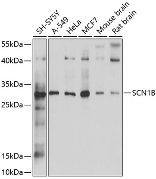
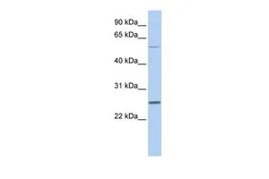
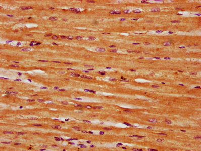
SCN1B Antibody (N-Terminus, Sepharose)
LS-C172071
ApplicationsImmunoPrecipitation
Product group Antibodies
TargetSCN1B
Overview
- SupplierLifeSpan BioSciences
- Product NameSCN1B Antibody (N-Terminus, Sepharose)
- Delivery Days Customer23
- ApplicationsImmunoPrecipitation
- Applications SupplierIP
- CertificationResearch Use Only
- ClonalityPolyclonal
- ConjugateSepharose
- Estimated Purity...
- Gene ID6324
- Target nameSCN1B
- Target descriptionsodium voltage-gated channel beta subunit 1
- Target synonymsATFB13; BRGDA5; DEE52; EIEE52; GEFSP1; sodium channel subunit beta-1; sodium channel, voltage gated, type I beta subunit; sodium channel, voltage-gated, type I, beta
- HostRabbit
- Storage Instruction2°C to 8°C
- UNSPSC12352203
Related products
Product group Antibodies
SCN1B Polyclonal AntibodyBS-6687R
ApplicationsImmunoFluorescence, ELISA, ImmunoCytoChemistry, ImmunoHistoChemistry, ImmunoHistoChemistry Frozen, ImmunoHistoChemistry Paraffin
TargetSCN1B
- SizePrice
Product group Antibodies
Anti-SCN1B AntibodyA12779
ApplicationsWestern Blot, ImmunoHistoChemistry
- SizePrice

Product group Antibodies
Anti-SCN1B Antibody Picoband(r)A03061-1-CARRIER-FREE
ApplicationsWestern Blot, ELISA, ImmunoHistoChemistry
TargetSCN1B
- SizePrice

Product group Antibodies
SCN1B antibody, InternalGTX47701
ApplicationsWestern Blot
TargetSCN1B
- SizePrice
Product group Antibodies
SCN1B AntibodyCSB-PA020835LA01HU
ApplicationsImmunoFluorescence, ELISA, ImmunoHistoChemistry
ReactivityHuman
TargetSCN1B
- SizePrice