

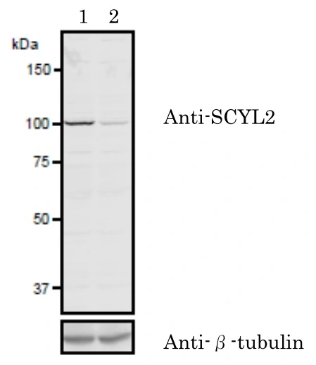
WB analysis of 293 cell lysate using GTX00732 SCYL2 antibody. Samples : Lane 1 : 293 cells transfected with control siRNA
Lane 2 : 293 cells transfected with si-SCYL2


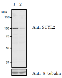
WB analysis of 293 cell lysate using GTX00732 SCYL2 antibody. Samples : Lane 1 : 293 cells transfected with control siRNA
Lane 2 : 293 cells transfected with si-SCYL2
SCYL2 antibody
GTX00732
ApplicationsImmunoFluorescence, ImmunoPrecipitation, Western Blot, ImmunoCytoChemistry
Product group Antibodies
ReactivityHamster, Human, Mouse, Rat
TargetSCYL2
Overview
- SupplierGeneTex
- Product NameSCYL2 antibody
- Delivery Days Customer9
- Application Supplier NoteWB: 1:1000. ICC/IF: 1:200-1:1000. IP: 1:200-1:1000. *Optimal dilutions/concentrations should be determined by the researcher.Not tested in other applications.
- ApplicationsImmunoFluorescence, ImmunoPrecipitation, Western Blot, ImmunoCytoChemistry
- CertificationResearch Use Only
- ClonalityPolyclonal
- Concentration1 mg/ml
- ConjugateUnconjugated
- Gene ID55681
- Target nameSCYL2
- Target descriptionSCY1 like pseudokinase 2
- Target synonymsAMC4, AMCNACC, CVAK104, SCY1-like protein 2, SCY1-like 2, SCY1-like, kinase-like 2, coated vesicle-associated kinase of 104 kDa
- HostRabbit
- IsotypeIgG
- Protein IDQ6P3W7
- Protein NameSCY1-like protein 2
- Scientific DescriptionThe protein encoded by this gene associates with clathrin-coated complexes at the plasma membrane and with endocytic coated vesicles. The encoded protein phosphorylates the beta2 subunit of the plasma membrane adapter complex AP2 and interacts with clathrin, showing involvement in clathrin-dependent pathways between the trans-Golgi network and the endosomal system. In addition, this protein has a role in the Wnt signaling pathway by targeting frizzled 5 (Fzd5) for lysosomal degradation. Two transcript variants encoding the same protein have been found for this gene. [provided by RefSeq, Dec 2015]
- ReactivityHamster, Human, Mouse, Rat
- Storage Instruction-20°C or -80°C,2°C to 8°C
- UNSPSC41116161
Datasheet
Related products
Product group Antibodies
SCYL2 AntibodyCSB-PA020874GA01HU
ApplicationsWestern Blot, ELISA
ReactivityHuman, Mouse, Rat
TargetSCYL2
- SizePrice
Product group Antibodies
Anti-SCYL2 AntibodyA101738
ApplicationsImmunoFluorescence, ImmunoPrecipitation, Western Blot
ReactivityHamster, Human, Mouse, Rat
- SizePrice

Product group Antibodies
Anti-SCYL2 Antibody Picoband(r)A08578-1-CARRIER-FREE
ApplicationsFlow Cytometry, ImmunoFluorescence, Western Blot, ELISA, ImmunoCytoChemistry
ReactivityHuman
TargetSCYL2
- SizePrice
Product group Antibodies

SCYL2 AntibodyLS-C748239
ApplicationsWestern Blot
ReactivityHuman, Mouse, Rat
TargetSCYL2
- SizePrice

Product group Antibodies
Anti-SCYL2 AntibodyHPA021050
ApplicationsImmunoHistoChemistry
ReactivityHuman
TargetSCYL2
- SizePrice
Product group Antibodies
SCYL2 antibodyGTX66174
ApplicationsImmunoFluorescence, Western Blot, ImmunoCytoChemistry
ReactivityHuman, Mouse, Rat
TargetSCYL2
- SizePrice

Product group Antibodies
SCYL2 antibodyGTX66175
ApplicationsWestern Blot
ReactivityHuman, Rat
TargetSCYL2
- SizePrice
Product group Antibodies
Anti-SCYL2 Antibody144-60223
ApplicationsWestern Blot
ReactivityHuman, Mouse, Rat
TargetSCYL2
- SizePrice