

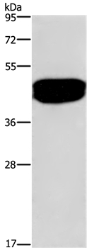
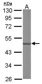
Western Blot analysis of Human prostate tissue using SDCCAG3 Polyclonal Antibody at dilution of 1:500
Western Blot analysis of Human prostate tissue using SDCCAG3 Polyclonal Antibody at dilution of 1:500
SDCCAG3 Polyclonal Antibody
E-AB-10275
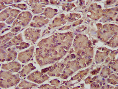
ApplicationsWestern Blot, ImmunoHistoChemistry
Product group Antibodies
TargetENTR1
Overview
- SupplierElabscience
- Product NameSDCCAG3 Polyclonal Antibody
- Delivery Days Customer12
- ApplicationsWestern Blot, ImmunoHistoChemistry
- Applications SupplierELISA WB IHC
- CertificationResearch Use Only
- ClonalityPolyclonal
- Concentration0.5 mg/ml
- ConjugateUnconjugated
- Gene ID10807
- Target nameENTR1
- Target descriptionendosome associated trafficking regulator 1
- Target synonymsNY-CO-3, SDCCAG3, SDDAG3, endosome-associated-trafficking regulator 1, antigen NY-CO-3, serologically defined colon cancer antigen 3
- HostRabbit
- IsotypeIgG
- Protein IDQ96C92
- Protein NameEndosome-associated-trafficking regulator 1
- Scientific DescriptionSDCCAG3 protein contains the region similar to the coiled-coil domain of the myosin tail. The same domain is present in the proteins related to the organelles/proteins trafficking, such as kinesin, Golgin-160, and dynein. SDCCAG3 function might be related to protein trafficking and secretion, May be involved in modulation of TNF response, May be involved in presentation of TNFRSF1A on the cell surface. Interaction of SDCCAG3 with the Arf GTPase activating protein GIT1 (G protein-coupled receptor kinase interactor-1). Overexpression of an ArfGAP-negative version of GIT1 also results in an increased number of multinucleate cells suggesting regulation of Arf-mediated vesicular trafficking or signaling via SDCCAG3.
- Storage Instruction-20°C
- UNSPSC41116161
MSDS
Related products
Product group Antibodies
ENTR1 AntibodyCSB-PA020897LA01HU
ApplicationsImmunoFluorescence, ELISA, ImmunoHistoChemistry
ReactivityHuman
TargetENTR1
- SizePrice

Product group Antibodies
Anti-ENTR1 Antibody Picoband(r)A31716-1-CARRIER-FREE
ApplicationsFlow Cytometry, Western Blot, ELISA
ReactivityHuman
TargetENTR1
- SizePrice

Product group Antibodies
Anti-SDCCAG3 AntibodyHPA029303
ApplicationsImmunoHistoChemistry
ReactivityHuman
TargetENTR1
- SizePrice
Product group Antibodies

SDCCAG3 AntibodyLS-C400701
ApplicationsWestern Blot, ELISA, ImmunoHistoChemistry
ReactivityHuman, Mouse
TargetENTR1
- SizePrice
Product group Antibodies
SDCCAG3 antibody [C1C3]GTX106180
ApplicationsWestern Blot
ReactivityHuman, Mouse
TargetENTR1
- SizePrice
Product group Antibodies
Anti-SDCCAG3 Antibody107-10586
ApplicationsWestern Blot
ReactivityHuman, Mouse
TargetENTR1
- SizePrice