Bio-Connect

Search results: 10-1141-01

565 to 576 of 3.214 products
Sort by
Product group Antibodies
LifeSpan BioSciences
ApplicationsImmunoFluorescence, Western Blot, ImmunoCytoChemistry
ReactivityOther Species
  • SizePrice
Western blot analysis of Paxillin using anti-Paxillin antibody (MA1080).
Product group Antibodies
Boster Bio
ApplicationsWestern Blot, ImmunoCytoChemistry
ReactivityBovine, Canine, Chicken, Equine, Hamster, Human, Mouse, Rabbit, Rat
TargetPXN
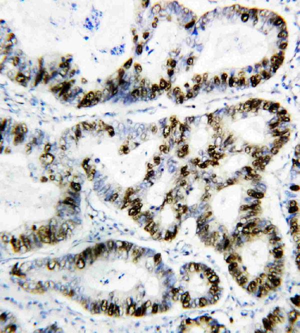
IHC analysis of PCNA using anti-PCNA antibody (MA1083). PCNA was detected in paraffin-embedded section of human Rectal cancer tissues. Heat mediated antigen retrieval was performed in citrate buffer (pH6, epitope retrieval solution) for 20 mins. The tissue section was blocked with 10% goat serum. The tissue section was then incubated with 1μg/ml mouse anti-PCNA Antibody (MA1083) overnight at 4°C. Biotinylated goat anti-mouse IgG was used as secondary antibody and incubated for 30 minutes at 37°C. The tissue section was developed using Strepavidin-Biotin-Complex (SABC)(Catalog # SA1021) with DAB as the chromogen.
Product group Antibodies
References
Boster Bio
ApplicationsWestern Blot, ImmunoCytoChemistry, ImmunoHistoChemistry
ReactivityBovine, Human, Mouse, Rat
TargetPCNA
Immunofluorescent analysis of Jurkat cells treated with Pervanadate, using Phospho-JAK2 (Y1007 + Y1008) Antibody.
Product group Antibodies
Boster Bio
ApplicationsFlow Cytometry, ImmunoFluorescence, ImmunoPrecipitation, Western Blot, ImmunoCytoChemistry, ImmunoHistoChemistry
ReactivityHuman, Mouse, Rat
TargetJAK2
Western blot analysis of Phospho-c-Jun (T91) expression in human brain lysate (P02038-1). Electrophoresis was performed on a 5-20% SDS-PAGE gel at 70V (Stacking gel) / 90V (Resolving gel) for 2-3 hours. The sample well of each lane was loaded with 50ug of sample under reducing conditions. After Electrophoresis, proteins were transferred to a Nitrocellulose membrane at 150mA for 50-90 minutes. Blocked the membrane with 5% Non-fat Milk/ TBS for 1.5 hour at RT. The membrane was incubated with rabbit anti-JUN monoclonal antibody (Catalog # P02038-1) overnight at 4 then washed with TBS-0.1%Tween 3 times with 5 minutes each and probed with a goat anti-rabbit IgG-HRP secondary antibody at a dilution of 1:10000 for 1.5 hour at RT. The signal is developed using an Enhanced Chemiluminescent detection (ECL) kit (Catalog # EK1002) with Tanon 5200 system. A specific band was detected for JUN
Product group Antibodies
Boster Bio
ApplicationsImmunoPrecipitation, Western Blot
ReactivityHuman, Mouse, Rat
TargetJUN
Western blot analysis of Phospho-c-Jun (S63) expression in NIH/3T3 cell lysate treated with Anisomycin (P02038). Electrophoresis was performed on a 5-20% SDS-PAGE gel at 70V (Stacking gel) / 90V (Resolving gel) for 2-3 hours. The sample well of each lane was loaded with 50ug of sample under reducing conditions. After Electrophoresis, proteins were transferred to a Nitrocellulose membrane at 150mA for 50-90 minutes. Blocked the membrane with 5% Non-fat Milk/ TBS for 1.5 hour at RT. The membrane was incubated with rabbit anti-Phospho-c-Jun (S63) monoclonal antibody (Catalog # P02038) overnight at 4°C, then washed with TBS-0.1%Tween 3 times with 5 minutes each and probed with a goat anti-rabbit IgG-HRP secondary antibody at a dilution of 1:10000 for 1.5 hour at RT. The signal is developed using an Enhanced Chemiluminescent detection (ECL) kit (Catalog # EK1002) with Tanon 5200 system.
Product group Antibodies
Boster Bio
ApplicationsImmunoFluorescence, Western Blot, ImmunoCytoChemistry, ImmunoHistoChemistry
ReactivityHuman, Mouse, Rat
TargetJUN
Western blot analysis of Phospho-JunD (S255) expression in (1) HeLa cell lysate treated with Alkaline Phosphatase; (2) HeLa cell lysate (P05609). Electrophoresis was performed on a 5-20% SDS-PAGE gel at 70V (Stacking gel) / 90V (Resolving gel) for 2-3 hours. The sample well of each lane was loaded with 50ug of sample under reducing conditions. After Electrophoresis, proteins were transferred to a Nitrocellulose membrane at 150mA for 50-90 minutes. Blocked the membrane with 5% Non-fat Milk/ TBS for 1.5 hour at RT. The membrane was incubated with rabbit anti-JUND monoclonal antibody (Catalog # P05609) overnight at 4 then washed with TBS-0.1%Tween 3 times with 5 minutes each and probed with a goat anti-rabbit IgG-HRP secondary antibody at a dilution of 1:10000 for 1.5 hour at RT. The signal is developed using an Enhanced Chemiluminescent detection (ECL) kit (Catalog # EK1002) with Tanon 5200 system. A specific band was detected for JUND
Product group Antibodies
Boster Bio
ApplicationsImmunoPrecipitation, Western Blot
ReactivityHuman, Mouse, Rat
TargetJUND
Product group Antibodies
References
Nordic-MUbio
ApplicationsELISA, ImmunoHistoChemistry
ReactivityHuman
Product group Antibodies
LifeSpan BioSciences
ApplicationsImmunoFluorescence, Western Blot, ImmunoHistoChemistry
ReactivityHuman, Mouse, Rat
  • SizePrice
Product group Antibodies
AdipoGen Life Sciences
ApplicationsFlow Cytometry
ReactivityMouse
TargetTnfrsf13b
  • SizePrice
Product group Antibodies
AdipoGen Life Sciences
ApplicationsFlow Cytometry
ReactivityMouse
TargetTnfrsf13b
  • SizePrice
Product group Antibodies
Absolute Antibody
ApplicationsELISA, RadioImmunoAssay
ReactivityPlant
  • SizePrice