

Search results: BIM
Product group Assays
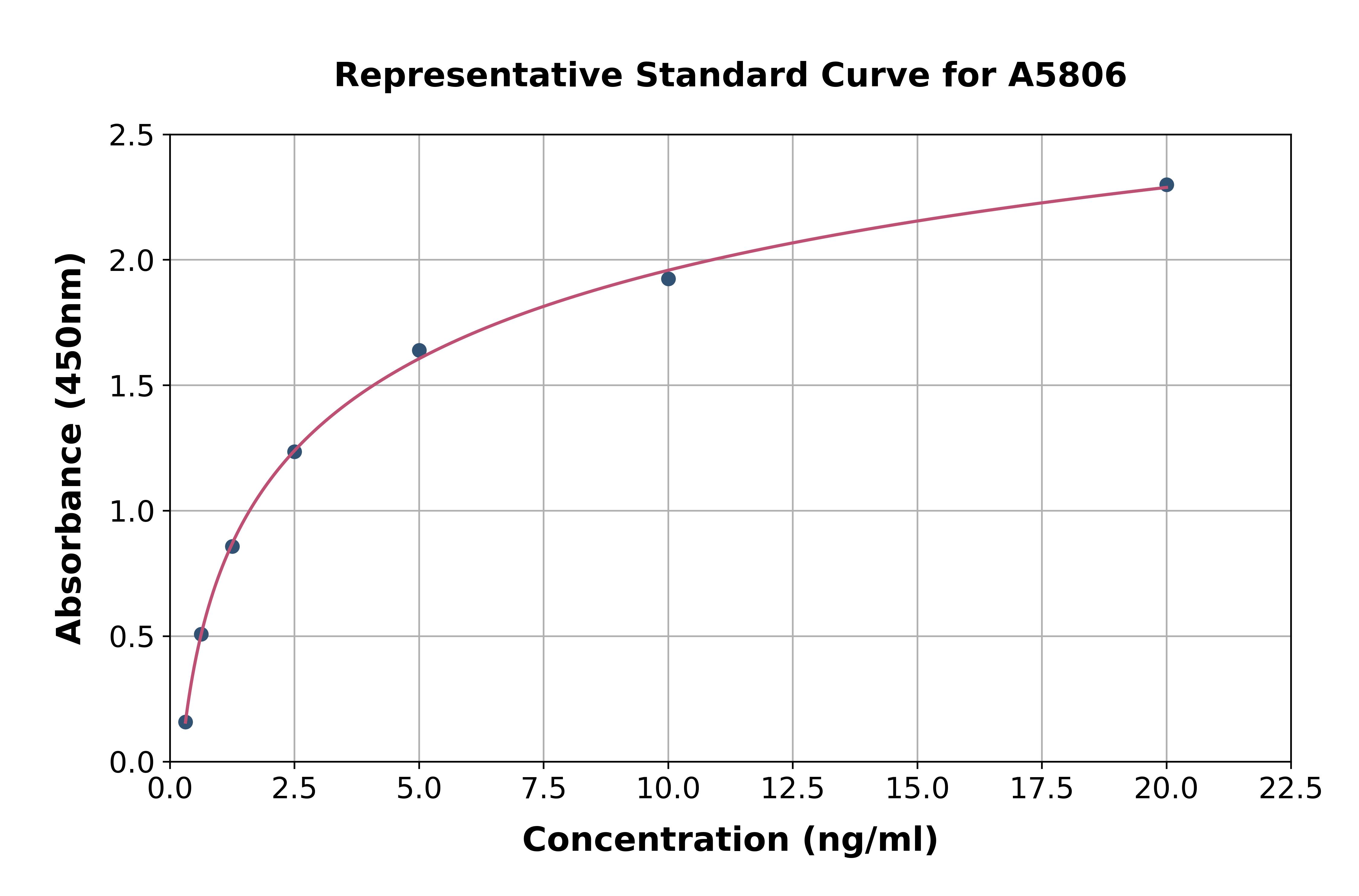
Human Bim ELISA KitA5806
Assay Sample TypeTissue homogenates, cell lysates and other biological fluids.
ReactivityHuman
- SizePrice
Product group Antibodies

Bim BH3 Domain AntibodyABX027202
ApplicationsWestern Blot, ELISA, ImmunoHistoChemistry
- SizePrice
Product group Antibodies
Anti- mouse Bim [ham151-149]AB03542-1.1-BT
ApplicationsFlow Cytometry, ImmunoPrecipitation, Western Blot
ReactivityMouse
TargetBcl2l11
- SizePrice
Product group Antibodies
Anti- mouse Bim [ham151-149]AB03542-23.0-BT
ApplicationsFlow Cytometry, ImmunoPrecipitation, Western Blot
ReactivityMouse
TargetBcl2l11
- SizePrice
Product group Antibodies
ApplicationsImmunoHistoChemistry, ImmunoHistoChemistry Paraffin
ReactivityHuman
TargetBcl2l11
- SizePrice

Product group Antibodies
Anti-Bim/Bcl2l11 Antibody Picoband(r)A01552-5-10UG
ApplicationsFlow Cytometry, Western Blot, ELISA
ReactivityMouse
TargetBcl2l11
- SizePrice
Product group Antibodies
Anti-Bim/Bcl2l11 Antibody Picoband(r)A01552-5-APC
ApplicationsFlow Cytometry, Western Blot, ELISA
ReactivityMouse
TargetBcl2l11
- SizePrice
Product group Antibodies
Anti-Bim/Bcl2l11 Antibody Picoband(r)A01552-5-BIOTIN
ApplicationsFlow Cytometry, Western Blot, ELISA
ReactivityMouse
TargetBcl2l11
- SizePrice
Product group Antibodies
Anti-Bim/Bcl2l11 Antibody Picoband(r)A01552-5-CARRIER-FREE
ApplicationsFlow Cytometry, Western Blot, ELISA
ReactivityMouse
TargetBcl2l11
- SizePrice
Product group Antibodies
Anti-Bim/Bcl2l11 Antibody Picoband(r)A01552-5-CY3
ApplicationsFlow Cytometry, Western Blot, ELISA
ReactivityMouse
TargetBcl2l11
- SizePrice
Didn't find what you were looking for?
Search through our product groups to find the right product
Back to overview