

Search results: CSB-E04595r
Product group Antibodies
CSB Rabbit pAb, BF594 conjugatedORB2518155
ApplicationsImmunoFluorescence
ReactivityHuman, Mouse, Rat
- SizePrice
Product group Antibodies
CSB Rabbit pAb, BF555 conjugatedORB2518156
ApplicationsImmunoFluorescence
ReactivityHuman, Mouse, Rat
- SizePrice
Product group Antibodies
CSB Rabbit pAb, BF488 conjugatedORB2518157
ApplicationsImmunoFluorescence
ReactivityHuman, Mouse, Rat
- SizePrice
Product group Antibodies
CSB Rabbit pAb, BF405 conjugatedORB2518158
ApplicationsImmunoFluorescence
ReactivityHuman, Mouse, Rat
- SizePrice
Product group Antibodies
CSB Rabbit pAb, BF350 conjugatedORB2518159
ApplicationsImmunoFluorescence
ReactivityHuman, Mouse, Rat
- SizePrice
Product group Antibodies
CSB Rabbit pAb, HRP conjugatedORB471604
ApplicationsImmunoHistoChemistry, ImmunoHistoChemistry Frozen, ImmunoHistoChemistry Paraffin
ReactivityHuman, Mouse, Rat
- SizePrice
Product group Antibodies
CSB Rabbit pAb, Cy5 conjugatedORB940025
ApplicationsImmunoFluorescence
ReactivityHuman, Mouse, Rat
- SizePrice
Product group Antibodies
CSB Rabbit pAb, Cy3 conjugatedORB985296
ApplicationsImmunoFluorescence
ReactivityHuman, Mouse, Rat
- SizePrice
Product group Antibodies
CSB Rabbit pAb, APC conjugatedORB996994
ApplicationsImmunoFluorescence
ReactivityHuman, Mouse, Rat
- SizePrice
Product group Antibodies
CSB Rabbit pAb, FITC conjugatedORB187089
ApplicationsImmunoFluorescence
ReactivityHuman, Mouse, Rat
- SizePrice
![CSB antibody [N2C1], Internal detects CSB protein by western blot analysis. A. 30 ug 294T whole cell lysate/extract. B. 30 ug A432 whole cell lysate/extract. C. 31 ug HeLa whole cell lysate/extract. D. 30 ug HepG3 whole cell lysate/extract. 5 % SDS-PAGE.](https://cdn.origene.com/assets/images/antibody/106/gtx104589-1-w.jpg)
Product group Antibodies
ApplicationsImmunoFluorescence, Western Blot, ImmunoHistoChemistry
ReactivityHuman
TargetERCC6
- SizePrice
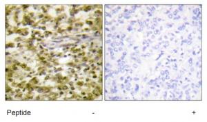
Product group Antibodies
ApplicationsImmunoHistoChemistry
ReactivityHuman
TargetERCC6
- SizePrice
Didn't find what you were looking for?
Search through our product groups to find the right product
Back to overview