

Search results: CSB-E04595r
Product group Antibodies
ApplicationsWestern Blot
ReactivityHuman
TargetERCC6
- SizePrice
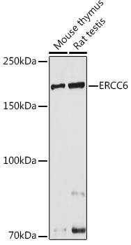
Product group Antibodies
ApplicationsWestern Blot, ImmunoHistoChemistry
ReactivityHuman, Mouse, Rat
TargetERCC6
- SizePrice
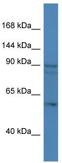
Product group Antibodies
ApplicationsWestern Blot
ReactivityBovine, Canine, Human, Mouse, Porcine, Rat
TargetERCC6
- SizePrice
Product group Genome Editing and Engineering
CSB CRISPR Activation Plasmid (h2)SC-401287-ACT-2
- SizePrice
Product group Genome Editing and Engineering
CSB Lentiviral Activation Particles (h2)SC-401287-LAC-2
- SizePrice
Product group Genome Editing and Engineering
CSB Lentiviral Activation Particles (h)SC-401287-LAC
- SizePrice
Product group DNA / RNA / Vectors
CSB shRNA (m) Lentiviral ParticlesSC-142603-V
CategoryshRNA
- SizePrice
Product group Genome Editing and Engineering
CSB CRISPR Activation Plasmid (m2)SC-435693-ACT-2
- SizePrice
Didn't find what you were looking for?
Search through our product groups to find the right product
Back to overview