

Search results: GCLM

Product group Antibodies
Anti-GCLM Antibody Picoband(r)A02948-2-PE
ApplicationsWestern Blot, ELISA
ReactivityHuman, Mouse, Rat
TargetGCLM
- SizePrice
Product group Antibodies
References
Anti-GCLM Antibody Picoband(r)A02948-2
ApplicationsWestern Blot, ELISA
ReactivityHuman, Mouse, Rat
TargetGCLM
- SizePrice

Product group Antibodies
Anti-GCLM Monoclonal AntibodyM02948-30UL
ApplicationsFlow Cytometry, ImmunoFluorescence, ImmunoPrecipitation, Western Blot, ImmunoCytoChemistry, ImmunoHistoChemistry
ReactivityHuman, Mouse, Rat
TargetGCLM
- SizePrice
Product group Antibodies
References
ApplicationsFlow Cytometry, ImmunoFluorescence, ImmunoPrecipitation, Western Blot, ImmunoCytoChemistry, ImmunoHistoChemistry
ReactivityHuman, Mouse, Rat
TargetGCLM
- SizePrice

Product group Assays
Mouse GCLM ELISA KitA7176
Assay Sample TypeSerum, plasma, tissue homogenates and other biological fluids.
ReactivityMouse
- SizePrice
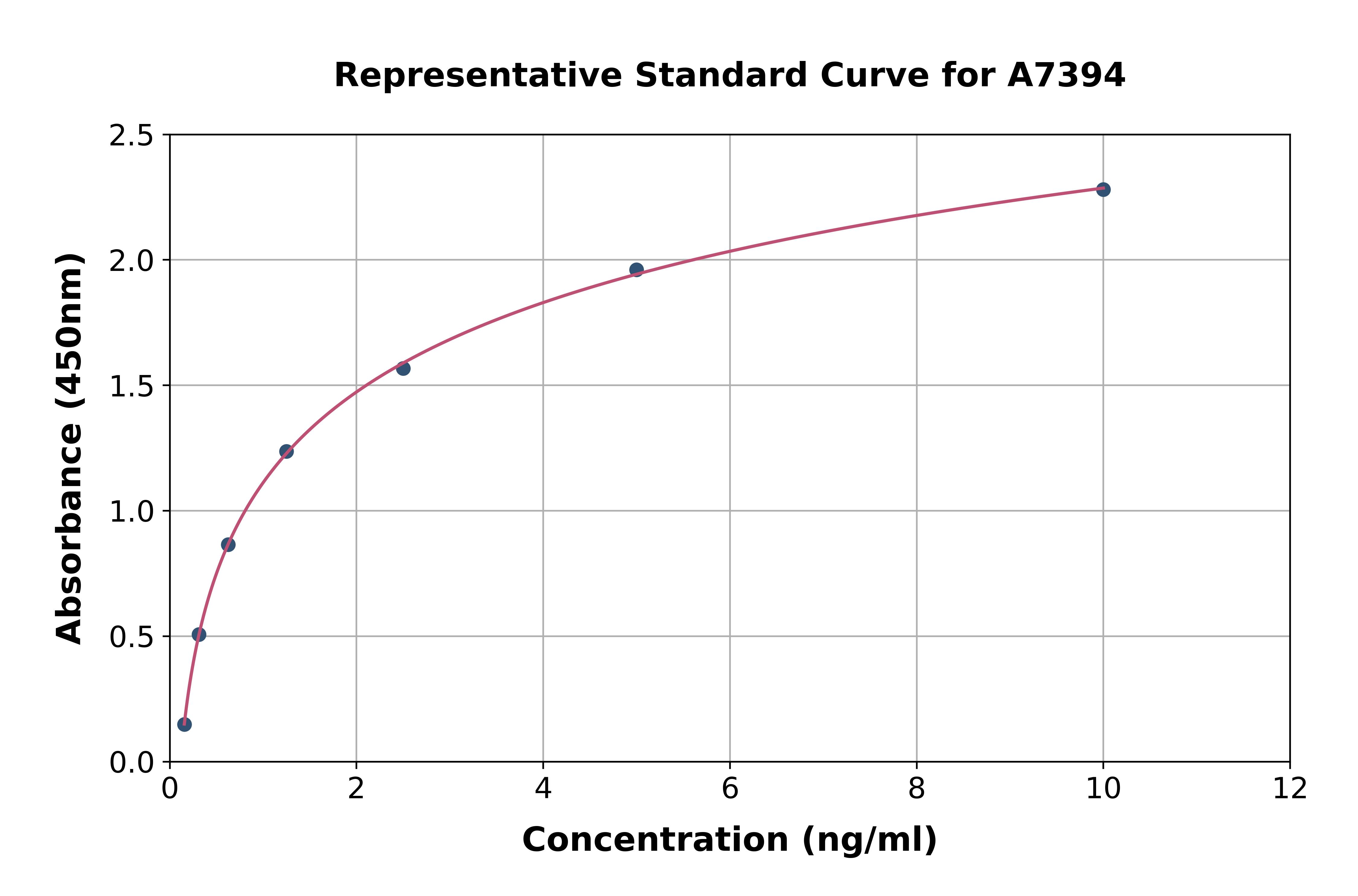
Product group Assays
Rat GCLM ELISA KitA7394
Assay Sample TypeTissue homogenates and other biological fluids.
ReactivityRat
- SizePrice
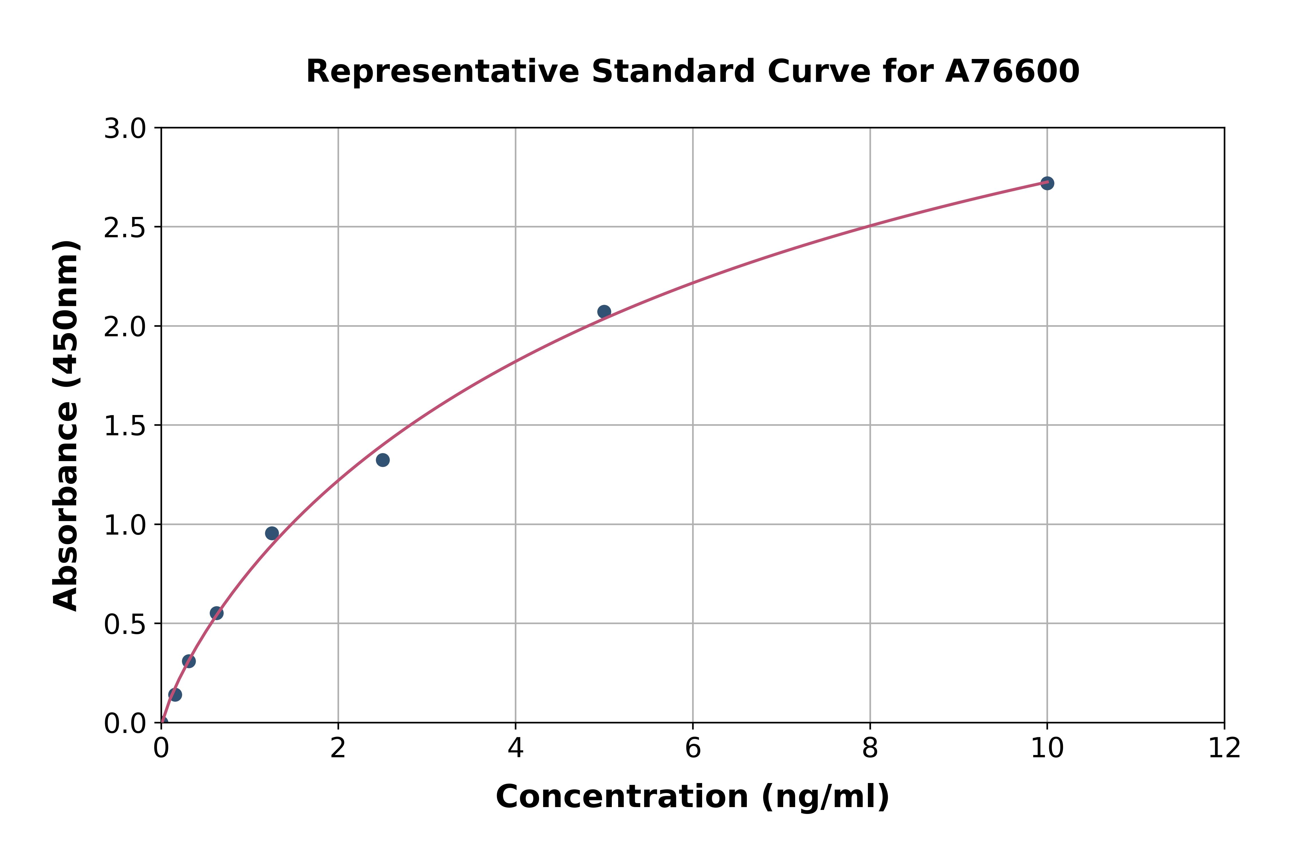
Product group Assays
Human GCLM ELISA KitA76600
Assay Sample TypePlasma, tissue homogenates and other biological fluids.
ReactivityHuman
- SizePrice

Product group Assays
Rat GCLM ELISA KitA79366
Assay Sample TypePlasma, tissue homogenates and other biological fluids.
ReactivityRat
- SizePrice
![Anti-GCLM Antibody [ARC0597]](https://www.antibodies.com/image/catalog/81/A81176_1.jpg)
Product group Antibodies
ApplicationsWestern Blot
ReactivityHuman
- SizePrice
Product group Antibodies

GCLM AntibodyLS-C806586
ApplicationsWestern Blot, ELISA, ImmunoHistoChemistry
ReactivityHuman, Mouse, Porcine, Rat
TargetGCLM
- SizePrice
Didn't find what you were looking for?
Search through our product groups to find the right product
Back to overview