

Search results: HEPACAM
Product group Proteins / Signaling Molecules
- SizePrice
Product group Proteins / Signaling Molecules
Protein IDQ14CZ8
- SizePrice
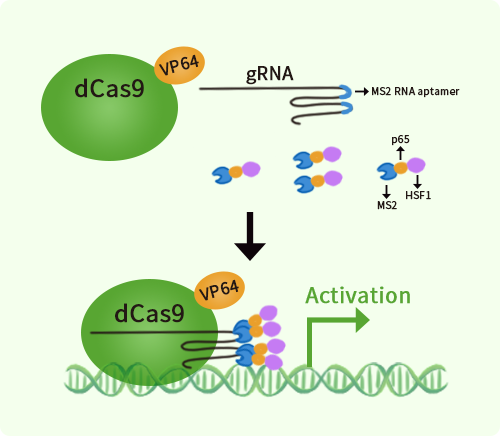
Product group Genome Editing and Engineering
- SizePrice
Product group DNA / RNA / Vectors
CategoryPlasmid / vector
- SizePrice
Product group Proteins / Signaling Molecules
- SizePrice

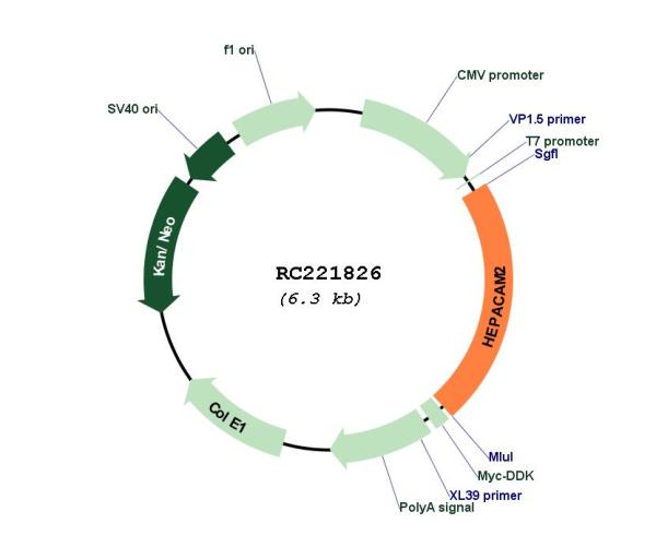
Product group DNA / RNA / Vectors
CategoryPlasmid / vector
- SizePrice
Product group DNA / RNA / Vectors
CategoryPlasmid / vector
- SizePrice
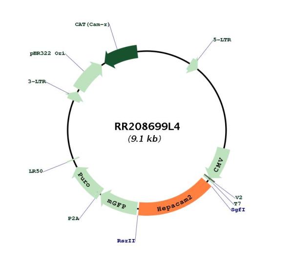
Product group DNA / RNA / Vectors
CategoryshRNA
- SizePrice
Product group DNA / RNA / Vectors
CategoryPlasmid / vector
- SizePrice
Product group DNA / RNA / Vectors
CategoryshRNA
- SizePrice
Product group Lysates / Extracts
- SizePrice
Product group DNA / RNA / Vectors
CategoryPlasmid / vector
- SizePrice
Didn't find what you were looking for?
Search through our product groups to find the right product
Back to overview