

Search results: MAP2K4 MEK4 MKK4 SEK1
Product group Antibodies
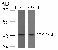
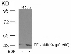
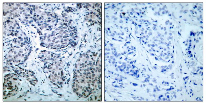
ApplicationsWestern Blot, ImmunoHistoChemistry
ReactivityHuman, Mouse, Rat
- SizePrice
Product group Antibodies
ApplicationsImmunoFluorescence, Western Blot, ImmunoHistoChemistry
ReactivityHuman, Mouse, Rat
- SizePrice
Product group Antibodies
ApplicationsImmunoFluorescence, ImmunoHistoChemistry
ReactivityHuman, Mouse, Rat
- SizePrice
Product group Assays
Assay Sample TypeResearchers plate their cell line of choice
ReactivityHuman, Mouse, Rat
- SizePrice
Product group Assays
Assay Sample TypeResearchers plate their cell line of choice
ReactivityHuman, Mouse, Rat
- SizePrice

Product group Antibodies
Anti-MEK4/MAP2K4 Antibody Picoband(r)A01725-1-10UG
ApplicationsWestern Blot, ELISA
ReactivityHuman, Mouse, Rat
TargetMAP2K4
- SizePrice
Product group Antibodies
Anti-MEK4/MAP2K4 Antibody Picoband(r)A01725-1-APC
ApplicationsWestern Blot, ELISA
ReactivityHuman, Mouse, Rat
TargetMAP2K4
- SizePrice
Product group Antibodies
Anti-MEK4/MAP2K4 Antibody Picoband(r)A01725-1-BIOTIN
ApplicationsWestern Blot, ELISA
ReactivityHuman, Mouse, Rat
TargetMAP2K4
- SizePrice
Product group Antibodies
Anti-MEK4/MAP2K4 Antibody Picoband(r)A01725-1-CARRIER-FREE
ApplicationsWestern Blot, ELISA
ReactivityHuman, Mouse, Rat
TargetMAP2K4
- SizePrice
Product group Antibodies
Anti-MEK4/MAP2K4 Antibody Picoband(r)A01725-1-CY3
ApplicationsWestern Blot, ELISA
ReactivityHuman, Mouse, Rat
TargetMAP2K4
- SizePrice
Product group Antibodies
Anti-MEK4/MAP2K4 Antibody Picoband(r)A01725-1-DYLIGHT488
ApplicationsWestern Blot, ELISA
ReactivityHuman, Mouse, Rat
TargetMAP2K4
- SizePrice
Product group Antibodies
Anti-MEK4/MAP2K4 Antibody Picoband(r)A01725-1-DYLIGHT550
ApplicationsWestern Blot, ELISA
ReactivityHuman, Mouse, Rat
TargetMAP2K4
- SizePrice
Didn't find what you were looking for?
Search through our product groups to find the right product
Back to overview