

Search results: MEK7 MKK7
Product group Antibodies
MEK7 Polyclonal Antibody, Cy5 ConjugatedBS-1979R-CY5
ApplicationsImmunoFluorescence, Western Blot, ImmunoCytoChemistry, ImmunoHistoChemistry, ImmunoHistoChemistry Frozen, ImmunoHistoChemistry Paraffin
ReactivityCanine, Equine, Human, Mouse, Porcine, Rat, Sheep
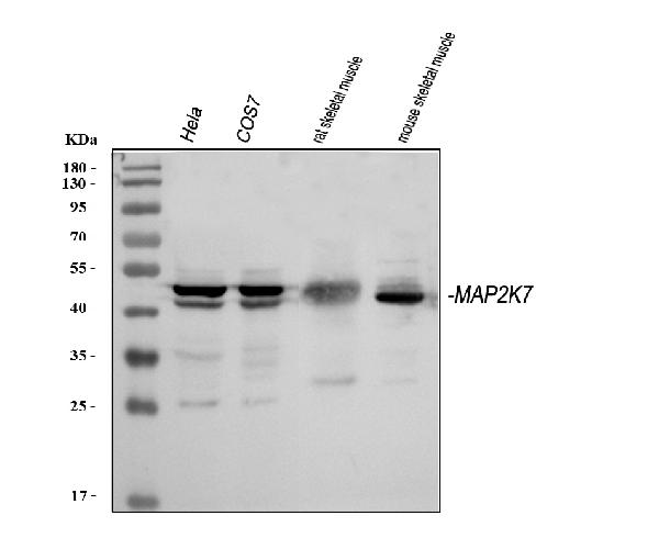
TargetMAP2K7
- SizePrice
Product group Antibodies
MEK7 Polyclonal Antibody, Cy7 ConjugatedBS-1979R-CY7
ApplicationsImmunoFluorescence, Western Blot, ImmunoCytoChemistry, ImmunoHistoChemistry, ImmunoHistoChemistry Frozen, ImmunoHistoChemistry Paraffin
ReactivityCanine, Equine, Human, Mouse, Porcine, Rat, Sheep
TargetMAP2K7
- SizePrice
Product group Antibodies
MEK7 Polyclonal Antibody, FITC ConjugatedBS-1979R-FITC
ApplicationsImmunoFluorescence, Western Blot, ImmunoCytoChemistry, ImmunoHistoChemistry, ImmunoHistoChemistry Frozen, ImmunoHistoChemistry Paraffin
ReactivityCanine, Equine, Human, Mouse, Porcine, Rat, Sheep
TargetMAP2K7
- SizePrice
Product group Antibodies
MEK7 Polyclonal Antibody, HRP ConjugatedBS-1979R-HRP
ApplicationsWestern Blot, ELISA, ImmunoHistoChemistry, ImmunoHistoChemistry Frozen, ImmunoHistoChemistry Paraffin
ReactivityCanine, Equine, Human, Mouse, Porcine, Rat, Sheep
TargetMAP2K7
- SizePrice
Product group Antibodies
MEK7 Polyclonal Antibody, PE Conjugatedbs-1979R-PE
ApplicationsImmunoFluorescence, Western Blot, ImmunoCytoChemistry, ImmunoHistoChemistry, ImmunoHistoChemistry Frozen, ImmunoHistoChemistry Paraffin
ReactivityCanine, Equine, Human, Mouse, Porcine, Rat, Sheep
TargetMAP2K7
- SizePrice
Product group Antibodies
MEK7 Polyclonal Antibody, RBITC ConjugatedBS-1979R-RBITC
ApplicationsImmunoFluorescence, Western Blot, ImmunoCytoChemistry, ImmunoHistoChemistry, ImmunoHistoChemistry Frozen, ImmunoHistoChemistry Paraffin
ReactivityCanine, Equine, Human, Mouse, Porcine, Rat, Sheep
TargetMAP2K7
- SizePrice
Product group Antibodies
Anti-MEK7/MAP2K7 Antibody Picoband(r)PA1922-10UG
ApplicationsFlow Cytometry, Western Blot
ReactivityChicken, Human, Monkey, Mouse, Rat
TargetMAP2K7
- SizePrice
Product group Antibodies
Anti-MEK7/MAP2K7 Antibody Picoband(r)PA1922-APC
ApplicationsFlow Cytometry, Western Blot
ReactivityChicken, Human, Monkey, Mouse, Rat
TargetMAP2K7
- SizePrice
Product group Antibodies
Anti-MEK7/MAP2K7 Antibody Picoband(r)PA1922-BIOTIN
ApplicationsFlow Cytometry, Western Blot
ReactivityChicken, Human, Monkey, Mouse, Rat
TargetMAP2K7
- SizePrice
Product group Antibodies
Anti-MEK7/MAP2K7 Antibody Picoband(r)PA1922-CARRIER-FREE
ApplicationsFlow Cytometry, Western Blot
ReactivityChicken, Human, Monkey, Mouse, Rat
TargetMAP2K7
- SizePrice
Product group Antibodies
Anti-MEK7/MAP2K7 Antibody Picoband(r)PA1922-CY3
ApplicationsFlow Cytometry, Western Blot
ReactivityChicken, Human, Monkey, Mouse, Rat
TargetMAP2K7
- SizePrice
Product group Antibodies
Anti-MEK7/MAP2K7 Antibody Picoband(r)PA1922-DYLIGHT488
ApplicationsFlow Cytometry, Western Blot
ReactivityChicken, Human, Monkey, Mouse, Rat
TargetMAP2K7
- SizePrice
Didn't find what you were looking for?
Search through our product groups to find the right product
Back to overview