

Search results: MEK7 MKK7
Product group Antibodies
Anti-MEK7/MAP2K7 Antibody Picoband(r)PB9764-DYLIGHT488
ApplicationsFlow Cytometry, ImmunoFluorescence, Western Blot, ImmunoCytoChemistry, ImmunoHistoChemistry
ReactivityHuman, Monkey, Mouse, Rat
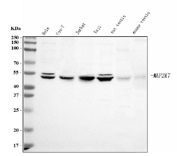
TargetMAP2K7
- SizePrice
Product group Antibodies
Anti-MEK7/MAP2K7 Antibody Picoband(r)PB9764-DYLIGHT550
ApplicationsFlow Cytometry, ImmunoFluorescence, Western Blot, ImmunoCytoChemistry, ImmunoHistoChemistry
ReactivityHuman, Monkey, Mouse, Rat
TargetMAP2K7
- SizePrice
Product group Antibodies
Anti-MEK7/MAP2K7 Antibody Picoband(r)PB9764-DYLIGHT594
ApplicationsFlow Cytometry, ImmunoFluorescence, Western Blot, ImmunoCytoChemistry, ImmunoHistoChemistry
ReactivityHuman, Monkey, Mouse, Rat
TargetMAP2K7
- SizePrice
Product group Antibodies
Anti-MEK7/MAP2K7 Antibody Picoband(r)PB9764-FITC
ApplicationsFlow Cytometry, ImmunoFluorescence, Western Blot, ImmunoCytoChemistry, ImmunoHistoChemistry
ReactivityHuman, Monkey, Mouse, Rat
TargetMAP2K7
- SizePrice
Product group Antibodies
Anti-MEK7/MAP2K7 Antibody Picoband(r)PB9764-HRP
ApplicationsFlow Cytometry, ImmunoFluorescence, Western Blot, ImmunoCytoChemistry, ImmunoHistoChemistry
ReactivityHuman, Monkey, Mouse, Rat
TargetMAP2K7
- SizePrice
Product group Antibodies
Anti-MEK7/MAP2K7 Antibody Picoband(r)PB9764-IFLUOR647
ApplicationsFlow Cytometry, ImmunoFluorescence, Western Blot, ImmunoCytoChemistry, ImmunoHistoChemistry
ReactivityHuman, Monkey, Mouse, Rat
TargetMAP2K7
- SizePrice
Product group Antibodies
ApplicationsFlow Cytometry, ImmunoFluorescence, Western Blot, ImmunoCytoChemistry, ImmunoHistoChemistry
ReactivityHuman, Monkey, Mouse, Rat
TargetMAP2K7
- SizePrice
Product group Antibodies
ApplicationsFlow Cytometry, ImmunoFluorescence, Western Blot, ImmunoCytoChemistry, ImmunoHistoChemistry
ReactivityHuman, Monkey, Mouse, Rat
TargetMAP2K7
- SizePrice
Product group Antibodies

MAP2K7 / MEK7 AntibodyLS-C16401
ApplicationsImmunoFluorescence, Western Blot, ELISA
ReactivityHuman, Mouse
TargetMAP2K7
- SizePrice
Product group Antibodies
MAP2K7 / MEK7 AntibodyLS-C768035
ApplicationsImmunoFluorescence, Western Blot
ReactivityHuman, Mouse
TargetMAP2K7
- SizePrice
Product group Antibodies
MAP2K7 / MEK7 AntibodyLS-C666781
ApplicationsWestern Blot, ELISA
ReactivityHuman
TargetMAP2K7
- SizePrice
Product group Antibodies
MAP2K7 / MEK7 AntibodyLS-C671543
ApplicationsImmunoFluorescence, ELISA
ReactivityHuman
TargetMAP2K7
- SizePrice
Didn't find what you were looking for?
Search through our product groups to find the right product
Back to overview