

Search results: NSP3
Product group Antibodies
- SizePrice
Product group Antibodies
- SizePrice
Product group Antibodies
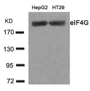
ApplicationsWestern Blot, ImmunoHistoChemistry, ImmunoHistoChemistry Paraffin
ReactivityHuman
TargetEIF4G1
- SizePrice
Product group Proteins / Signaling Molecules
PrEST Antigen SH2D3CAPREST75183
Protein IDQ8N5H7
- SizePrice
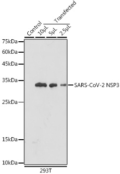
Product group Antibodies
ApplicationsWestern Blot
ReactivityVirus
TargetORF1ab
- SizePrice

Product group Antibodies
Anti-SH2D3C AntibodyHPA047586
ApplicationsWestern Blot, ImmunoHistoChemistry
ReactivityHuman
TargetSH2D3C
- SizePrice
Product group Antibodies
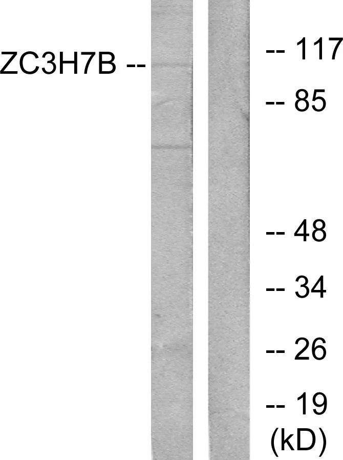
ZC3H7B antibodyGTX87217
ApplicationsWestern Blot
ReactivityHuman
TargetZC3H7B
- SizePrice
Product group Antibodies
EIF4G1 (Ab-1232) AntibodyCSB-PA061737
ApplicationsImmunoFluorescence, Western Blot, ELISA, ImmunoHistoChemistry
ReactivityHuman
TargetEIF4G1
- SizePrice
Product group Antibodies
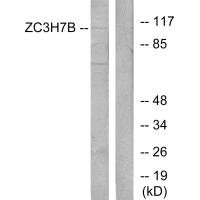
ZC3H7B AntibodyCSB-PA197208
ApplicationsWestern Blot, ELISA
ReactivityHuman
TargetZC3H7B
- SizePrice
Didn't find what you were looking for?
Search through our product groups to find the right product
Back to overview