

Search results: PDCD4

Product group Antibodies
Anti-PDCD4 Antibody Picoband(r)A01105-IFLUOR647
ApplicationsFlow Cytometry, Western Blot, ImmunoHistoChemistry
ReactivityHuman
TargetPDCD4
- SizePrice
Product group Antibodies
Anti-PDCD4 Antibody Picoband(r)A01105-PE
ApplicationsFlow Cytometry, Western Blot, ImmunoHistoChemistry
ReactivityHuman
TargetPDCD4
- SizePrice
Product group Antibodies
ApplicationsFlow Cytometry, Western Blot, ImmunoHistoChemistry
ReactivityHuman
TargetPDCD4
- SizePrice

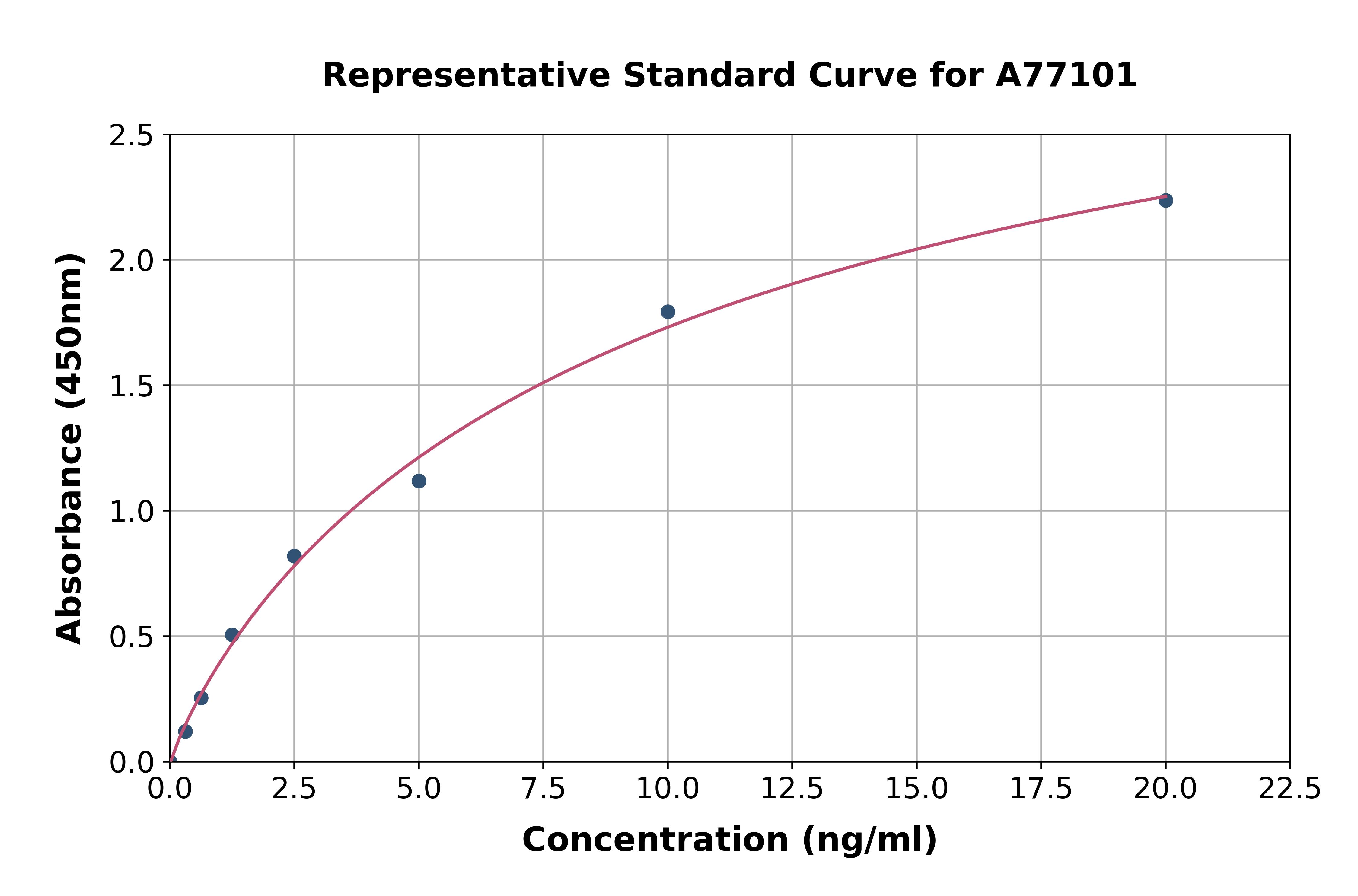
Product group Assays
Human PDCD4 ELISA KitA77101
Assay Sample TypePlasma, tissue homogenates and other biological fluids.
ReactivityHuman
- SizePrice
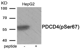
Product group Antibodies
Phospho-PDCD4 (Ser67) AntibodyCSB-PA047784
ApplicationsWestern Blot, ELISA
ReactivityHuman, Mouse, Rat
TargetPDCD4
- SizePrice

Product group Proteins / Signaling Molecules
Human PDCD4 Recombinant ProteinPROTQ53EL6
ApplicationsOther Application
Protein IDQ53EL6
- SizePrice
Product group Proteins / Signaling Molecules
Human PDCD4 Recombinant ProteinPROTQ53EL6
ApplicationsOther Application
Protein IDQ53EL6
- SizePrice
Didn't find what you were looking for?
Search through our product groups to find the right product
Back to overview