

Search results: Survivin
Product group Proteins / Signaling Molecules
Survivin peptideGTX27866
ApplicationsNeutralisation/Blocking
- SizePrice
Product group Proteins / Signaling Molecules
Survivin peptideGTX28122
ApplicationsNeutralisation/Blocking
- SizePrice
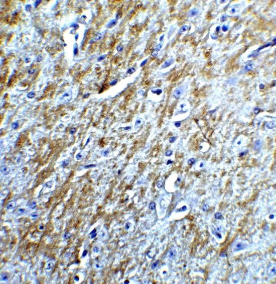
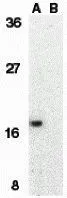
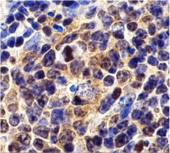
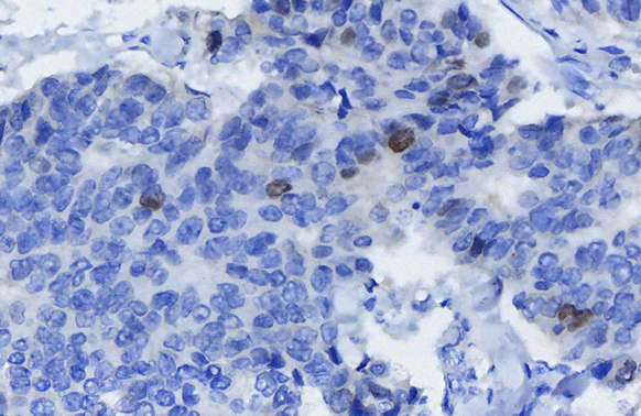
Product group Antibodies
Survivin antibodyGTX31651
ApplicationsImmunoFluorescence, Western Blot, ELISA, ImmunoCytoChemistry, ImmunoHistoChemistry, ImmunoHistoChemistry Paraffin
ReactivityHuman, Mouse
TargetBIRC5
- SizePrice
Product group Antibodies
Survivin antibodyGTX31652
ApplicationsImmunoFluorescence, Western Blot, ELISA, ImmunoCytoChemistry
ReactivityHuman
TargetBIRC5
- SizePrice
Product group Antibodies
Survivin antibodyGTX31653
ApplicationsWestern Blot, ELISA, ImmunoHistoChemistry, ImmunoHistoChemistry Paraffin
ReactivityMouse
TargetBIRC5
- SizePrice
Product group Antibodies
Survivin antibodyGTX100052
ApplicationsImmunoFluorescence, ImmunoPrecipitation, Western Blot, ImmunoCytoChemistry, ImmunoHistoChemistry, ImmunoHistoChemistry Paraffin
ReactivityHuman, Mouse
TargetBIRC5
- SizePrice
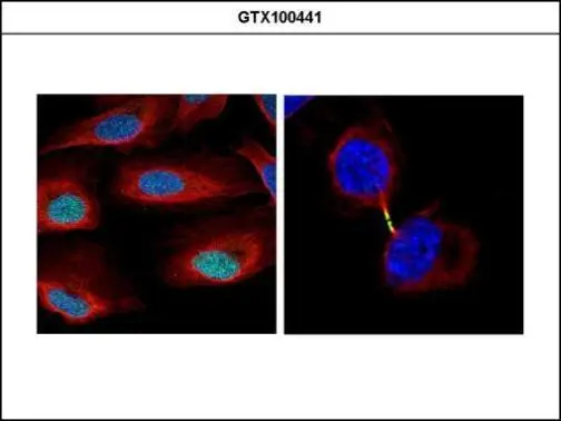
Product group Antibodies
Survivin antibodyGTX100441
ApplicationsImmunoFluorescence, ImmunoPrecipitation, Western Blot, ImmunoCytoChemistry, ImmunoHistoChemistry, ImmunoHistoChemistry Paraffin
ReactivityHuman, Mouse
TargetBIRC5
- SizePrice

Didn't find what you were looking for?
Search through our product groups to find the right product
Back to overview