

Search results: TCF3
Product group Antibodies
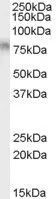
TCF3 antibody, InternalGTX89171
ApplicationsWestern Blot, ImmunoHistoChemistry, ImmunoHistoChemistry Paraffin
ReactivityHuman
- SizePrice
Product group Antibodies
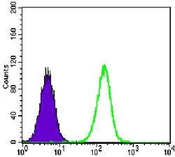
TCF3 antibody [6B8]GTX83355
ApplicationsFlow Cytometry, Western Blot, ELISA
ReactivityHuman
- SizePrice
Product group Antibodies
TCF3 / E2A AntibodyLS-C108663
ApplicationsImmunoFluorescence, Western Blot, ELISA, ImmunoHistoChemistry
ReactivityHuman, Mouse, Rat
- SizePrice
Product group Antibodies
TCF3 / E2A AntibodyLS-C802208
ApplicationsFlow Cytometry, Western Blot, ELISA
ReactivityHuman
- SizePrice
Product group Antibodies
TCF3 / E2A AntibodyLS-C822604
ApplicationsWestern Blot, ELISA, ImmunoHistoChemistry
ReactivityHuman, Mouse, Rat
- SizePrice

Product group Antibodies
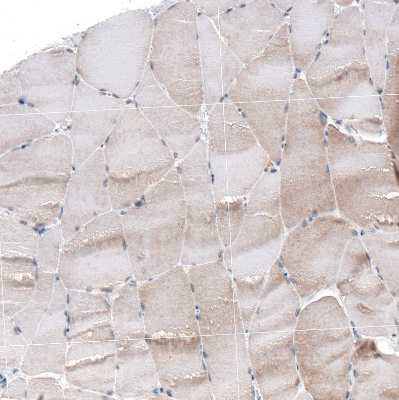
Anti-TCF3HPA062476
ApplicationsWestern Blot, ImmunoHistoChemistry
ReactivityHuman
TargetTCF3
- SizePrice
Product group Antibodies
Anti-TCF3HPA049808
ApplicationsImmunoCytoChemistry, ImmunoHistoChemistry
ReactivityHuman
TargetTCF3
- SizePrice
Product group Antibodies
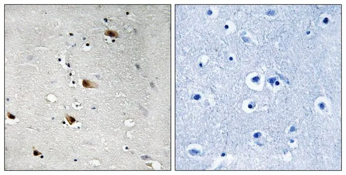
TCF3 (phospho Thr355) antibodyGTX55358
ApplicationsImmunoHistoChemistry, ImmunoHistoChemistry Paraffin
ReactivityHuman
- SizePrice
Product group Antibodies
ApplicationsWestern Blot, ELISA, ImmunoHistoChemistry
ReactivityHuman
- SizePrice

Product group Antibodies
TCF3 / E2A Antibody (aa51-100)LS-C206126
ApplicationsWestern Blot
ReactivityHuman
- SizePrice
Didn't find what you were looking for?
Search through our product groups to find the right product
Back to overview