

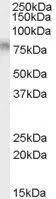
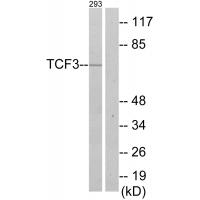
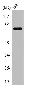
WB analysis of Daudi cell lysate using GTX89171 TCF3 / E2A antibody, Internal. Dilution : 0.01μg/ml Loading : 35μg protein in RIPA buffer

WB analysis of Daudi cell lysate using GTX89171 TCF3 / E2A antibody, Internal. Dilution : 0.01μg/ml Loading : 35μg protein in RIPA buffer
TCF3 / E2A antibody, Internal
GTX89171
ApplicationsWestern Blot, ImmunoHistoChemistry, ImmunoHistoChemistry Paraffin
Product group Antibodies
ReactivityHuman
TargetTCF3
Overview
- SupplierGeneTex
- Product NameTCF3 / E2A antibody, Internal
- Delivery Days Customer9
- Application Supplier NoteWB: 0.01-0.03microg/ml. IHC-P: 3-5microg/ml. *Optimal dilutions/concentrations should be determined by the researcher.Not tested in other applications.
- ApplicationsWestern Blot, ImmunoHistoChemistry, ImmunoHistoChemistry Paraffin
- CertificationResearch Use Only
- ClonalityPolyclonal
- Concentration0.50 mg/ml
- ConjugateUnconjugated
- Gene ID6929
- Target nameTCF3
- Target descriptiontranscription factor 3
- Target synonymsAGM8, AGM8A, AGM8B, E2A, E47, ITF1, TCF-3, VDIR, bHLHb21, p75, transcription factor E2-alpha, E2A-HLF fusion transcript protein, NOL1-TCF3 fusion, VDR interacting repressor, class B basic helix-loop-helix protein 21, helix-loop-helix protein HE47, immunoglobulin transcription factor 1, kappa-E2-binding factor, negative vitamin D response element-binding protein, transcription factor 3 (E2A immunoglobulin enhancer binding factors E12/E47), transcription factor ITF-1, vitamin D receptor-interacting repressor
- HostGoat
- IsotypeIgG
- Protein IDP15923
- Protein NameTranscription factor E2-alpha
- Scientific DescriptionThis gene encodes a member of the E protein (class I) family of helix-loop-helix transcription factors. E proteins activate transcription by binding to regulatory E-box sequences on target genes as heterodimers or homodimers, and are inhibited by heterodimerization with inhibitor of DNA-binding (class IV) helix-loop-helix proteins. E proteins play a critical role in lymphopoiesis, and the encoded protein is required for B and T lymphocyte development. Deletion of this gene or diminished activity of the encoded protein may play a role in lymphoid malignancies. This gene is also involved in several chromosomal translocations that are associated with lymphoid malignancies including pre-B-cell acute lymphoblastic leukemia (t(1;19), with PBX1), childhood leukemia (t(19;19), with TFPT) and acute leukemia (t(12;19), with ZNF384). Alternatively spliced transcript variants encoding multiple isoforms have been observed for this gene, and a pseudogene of this gene is located on the short arm of chromosome 9. [provided by RefSeq, Sep 2011]
- ReactivityHuman
- Storage Instruction-20°C or -80°C,2°C to 8°C
- UNSPSC41116161
Datasheet
Related products
Product group Antibodies
Anti-TCF3 AntibodyA38611
ApplicationsWestern Blot, ImmunoHistoChemistry
ReactivityHuman, Mouse, Rat
- SizePrice
Product group Antibodies
Anti-TCF3 Antibody101-11755
ApplicationsWestern Blot, ELISA
TargetTCF3
- SizePrice

Product group Antibodies
Anti-Phospho-E2A (T355) TCF3 AntibodyA00095T355
ApplicationsImmunoFluorescence, ELISA, ImmunoHistoChemistry
ReactivityHuman, Mouse, Rat
TargetTCF3
- SizePrice
Product group Antibodies
TCF3 Recombinant AntibodyBSM-60695R
ApplicationsImmunoFluorescence, Western Blot, ImmunoHistoChemistry, ImmunoHistoChemistry Frozen, ImmunoHistoChemistry Paraffin
ReactivityHuman
TargetTCF3
- SizePrice
Product group Antibodies
TCF3 AntibodyCSB-PA004248
ApplicationsWestern Blot, ELISA, ImmunoHistoChemistry
ReactivityHuman, Mouse, Rat
TargetTCF3
- SizePrice
Product group Antibodies
TCF3 / E2A antibodyGTX31402
ApplicationsWestern Blot, ELISA, ImmunoHistoChemistry, ImmunoHistoChemistry Paraffin
ReactivityHuman, Mouse, Rat
TargetTCF3
- SizePrice
Product group Antibodies

TCF3 / E2A AntibodyLS-C499896
ApplicationsELISA, ImmunoHistoChemistry
ReactivityHuman
TargetTCF3
- SizePrice
![FACS analysis of A549 cells using GTX83355 TCF3 / E2A antibody [6B8]. Green : TCF3 Purple : negative control](https://www.genetex.com/upload/website/prouct_img/normal/GTX83355/GTX83355_20170912_FACS_w_23061322_378.webp)
Product group Antibodies
TCF3 / E2A antibody [6B8]GTX83355
ApplicationsFlow Cytometry, Western Blot, ELISA
ReactivityHuman
TargetTCF3
- SizePrice

Product group Antibodies
Anti-TCF3 AntibodyHPA049808
ApplicationsChIP Chromatin ImmunoPrecipitation, ImmunoCytoChemistry, ImmunoHistoChemistry
ReactivityHuman
TargetTCF3
- SizePrice
Product group Antibodies
TCF3 / E2A antibodyGTX109885
ApplicationsImmunoFluorescence, ImmunoPrecipitation, Western Blot, ImmunoCytoChemistry, ImmunoHistoChemistry, ImmunoHistoChemistry Paraffin
ReactivityHuman, Mouse, Rat
TargetTCF3
- SizePrice