

Search results: akt
Product group Antibodies
Rabbit Polyclonal AntibodyTA392565
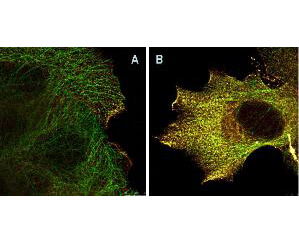
ApplicationsImmunoFluorescence, Western Blot
ReactivityHuman, Mouse, Rat
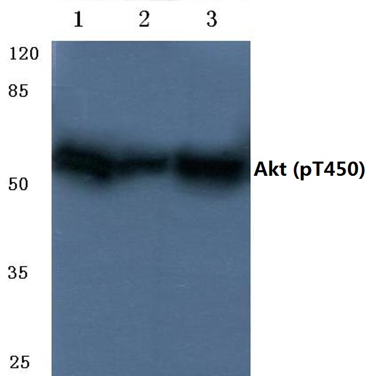
TargetAKT1
- SizePrice

Product group Antibodies
Rabbit Polyclonal AntibodyTA392568
ApplicationsWestern Blot
ReactivityHuman, Mouse, Rat
TargetAKT1
- SizePrice
Product group Antibodies
Rabbit Polyclonal AntibodyTA392572
ApplicationsImmunoFluorescence, Western Blot
ReactivityHuman, Mouse, Rat
TargetAKT1
- SizePrice
Product group Antibodies
Rabbit Polyclonal AntibodyTA392577
ApplicationsImmunoFluorescence, Western Blot
ReactivityHuman, Mouse, Rat
TargetAKT1
- SizePrice
Product group Antibodies
Rabbit Polyclonal AntibodyTA392582
ApplicationsImmunoFluorescence, Western Blot
ReactivityHuman, Mouse, Rat
TargetAKT1
- SizePrice
Product group Antibodies
Rabbit Polyclonal AntibodyTA392585
ApplicationsWestern Blot
ReactivityHuman, Mouse, Rat
TargetAKT1
- SizePrice
Product group Antibodies
Rabbit Polyclonal AntibodyTA392586
ApplicationsWestern Blot
ReactivityHuman, Mouse, Rat
TargetAKT1
- SizePrice
Product group Antibodies
Rabbit Polyclonal AntibodyTA392587
ApplicationsWestern Blot
ReactivityHuman, Mouse, Rat
TargetAKT1
- SizePrice
Product group Antibodies
Rabbit Polyclonal AntibodyTA392643
ApplicationsWestern Blot
ReactivityHuman, Mouse, Rat
TargetAKT1
- SizePrice
Product group Antibodies
Rabbit Polyclonal AntibodyTA392909
ApplicationsWestern Blot
ReactivityHuman, Mouse, Rat
TargetAKT1
- SizePrice
Product group Antibodies
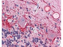
ApplicationsFlow Cytometry, ImmunoFluorescence, Western Blot, ELISA, ImmunoHistoChemistry
ReactivityHuman, Mouse
TargetAKT1
- SizePrice
Product group Antibodies
ApplicationsFlow Cytometry, Western Blot, ELISA, ImmunoHistoChemistry
ReactivityHuman, Mouse
TargetAKT1
- SizePrice
Didn't find what you were looking for?
Search through our product groups to find the right product
Back to overview