

Search results: anti-cea
Product group Antibodies
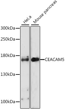
Anti-CEACAM5 AntibodyA88354
ApplicationsWestern Blot, ImmunoHistoChemistry
- SizePrice
Product group Antibodies
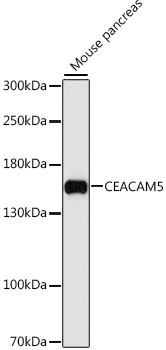
Anti-CEACAM5 AntibodyA36772
ApplicationsWestern Blot, ImmunoHistoChemistry
- SizePrice
Product group Antibodies
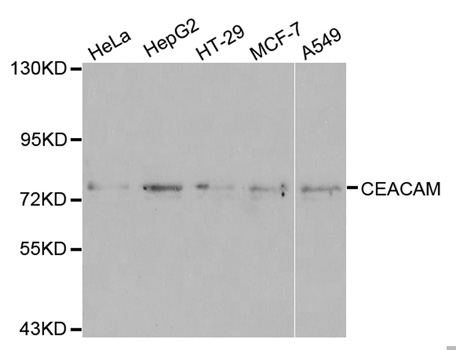
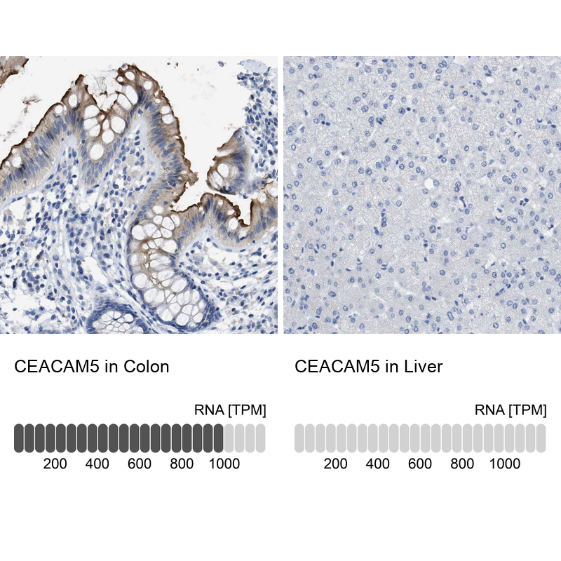
Anti-CEACAM5 AntibodyHPA019758
ApplicationsWestern Blot, ImmunoCytoChemistry, ImmunoHistoChemistry
ReactivityHuman
TargetCEACAM5
- SizePrice
Product group Antibodies

Anti-CEACAM5 [NbCEA5], Camelid VHH, His-tagged,AB04388-34.11-BS
ApplicationsFlow Cytometry, ELISA, Other Application
TargetCEACAM5
- SizePrice
Product group Antibodies
Anti-CEACAM5 [NbCEA5], Camelid VHH, His-tagged,AB04388-34.11-BT
ApplicationsFlow Cytometry, ELISA, Other Application
TargetCEACAM5
- SizePrice
Product group Antibodies
Anti-CEACAM5 [NbCEA5], Camelid VHH, His-tagged,AB04388-34.11
ApplicationsFlow Cytometry, ELISA, Other Application
TargetCEACAM5
- SizePrice
Product group Antibodies
ApplicationsWestern Blot, ELISA
- SizePrice
![Anti-CEACAM1 Antibody [ARC0649]](https://www.antibodies.com/image/catalog/81/A81072_1.jpg)
Product group Antibodies
ApplicationsWestern Blot, ImmunoHistoChemistry
- SizePrice

Product group Antibodies
Anti-CEACAM1 Antibody Picoband(r)A00923-2-BIOTIN
ApplicationsWestern Blot, ELISA
TargetCEACAM1
- SizePrice
Product group Antibodies
Anti-CEACAM1 Antibody Picoband(r)A00923-2-CARRIER-FREE
ApplicationsWestern Blot, ELISA
TargetCEACAM1
- SizePrice
Didn't find what you were looking for?
Search through our product groups to find the right product
Back to overview