

Search results: lacZ
Product group Antibodies
ApplicationsFlow Cytometry, ImmunoFluorescence, ImmunoPrecipitation, Western Blot, ELISA
- SizePrice
Product group Antibodies
lacZ Antibody, HRP conjugatedCSB-PA009476LB01ENV
ApplicationsELISA
ReactivityBacteria
- SizePrice
Product group Antibodies
lacZ Antibody, FITC conjugatedCSB-PA009476LC01ENV
ReactivityBacteria
- SizePrice
Product group Antibodies
lacZ Antibody, Biotin conjugatedCSB-PA009476LD01ENV
ApplicationsELISA
ReactivityBacteria
- SizePrice
Product group Antibodies
ApplicationsWestern Blot, ELISA, ImmunoHistoChemistry
ReactivityBacteria
TargetlacZ
- SizePrice
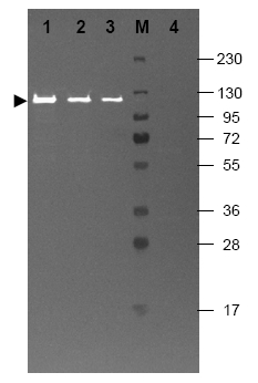
![Western blot using Beta-galactosidase antibody. shows detection of a band at ~117 kDa (lane 1) corresponding to the protein present in partially purified preparations. Approximately 50 ng of protein was separated on a 4-20% Tris-Glycine gel by SDSPAGE and transferred onto nitrocellulose. After blocking the membrane was probed with the primary antibody diluted to 1/1,000. Reaction occurred overnight at 4°C followed by washes and reaction with a 1/10,000 dilution of IRDye800 (TM) conjugated goat-a-rabbit IgG [H&L] for 45 min at RT (800 nm channel, green). Molecular weight estimation was made by comparison to prestained MW markers in lane M (700 nm channel, red). RDye800 (TM) fluorescence image was captured using the Odyssey (R) infrared imaging system developed by LI-COR. IRDye is a trademark of LI-COR, Inc. Other detection systems will yield similar results.](https://cdn.origene.com/assets/images/antibody/acris/r1064ps-1-w.jpg)
Product group Antibodies
ApplicationsImmunoFluorescence, ImmunoPrecipitation, Western Blot, ELISA, ImmunoHistoChemistry
ReactivityBacteria
TargetlacZ
- SizePrice
Product group Antibodies
lacZ Chicken Polyclonal AntibodyAP31768PU-N
ApplicationsImmunoFluorescence, Western Blot, ImmunoHistoChemistry
TargetlacZ
- SizePrice
Product group Antibodies
lacZ Rabbit Polyclonal AntibodyAP21258BT-N
ApplicationsImmunoDiffusion, ImmunoFluorescence, ImmunoPrecipitation, Western Blot, ELISA, RadioImmunoAssay
ReactivityBacteria
TargetlacZ
- SizePrice
Product group Antibodies
lacZ Rabbit Polyclonal AntibodyR1064HRPS
ApplicationsWestern Blot, ELISA, ImmunoHistoChemistry
ReactivityBacteria
TargetlacZ
- SizePrice
Product group Antibodies
ApplicationsWestern Blot, ELISA, ImmunoHistoChemistry
ReactivityBacteria
TargetlacZ
- SizePrice
Product group Antibodies
ApplicationsImmunoFluorescence, Western Blot
ReactivityBacteria
TargetlacZ
- SizePrice
Didn't find what you were looking for?
Search through our product groups to find the right product
Back to overview