Bio-Connect

Search results: p53 TP53

685 to 696 of 16.840 products
Sort by
Product group Antibodies
Cusabio
ApplicationsELISA
ReactivityHuman
TargetTP53TG5
  • SizePrice
Product group Antibodies
Cusabio
ReactivityHuman
TargetTP53TG5
  • SizePrice
Product group Antibodies
Cusabio
ApplicationsELISA
ReactivityHuman
TargetTP53TG5
  • SizePrice
IHC analysis of p53 using anti-p53 antibody (MA1078). p53 was detected in paraffin-embedded section of human mammary cancer tissues. Heat mediated antigen retrieval was performed in citrate buffer (pH6, epitope retrieval solution) for 20 mins. The tissue section was blocked with 10% goat serum. The tissue section was then incubated with 1μg/ml mouse anti-p53 Antibody (MA1078) overnight at 4°C. Biotinylated goat anti-mouse IgG was used as secondary antibody and incubated for 30 minutes at 37°C. The tissue section was developed using Strepavidin-Biotin-Complex (SABC)(Catalog # SA1021) with DAB as the chromogen.
Product group Antibodies
References
Boster Bio
ApplicationsWestern Blot, ImmunoCytoChemistry, ImmunoHistoChemistry
ReactivityHuman
TargetTP53
  • SizePrice
Product group Proteins / Signaling Molecules
GeneTex
ApplicationsNeutralisation/Blocking
  • SizePrice
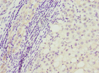
Immunohistochemistry of paraffin-embedded human liver cancer using CSB-PA884624DSR2HU at dilution of 1:100
Product group Antibodies
Cusabio
TP53AIP1 AntibodyCSB-PA884624DSR2HU
ApplicationsELISA, ImmunoHistoChemistry
ReactivityHuman
TargetTP53AIP1
  • SizePrice
Western blot analysis of p53 expression in (1) Raji cell lysate; (2) HepG2 cell lysate (M00001-2). Electrophoresis was performed on a 5-20% SDS-PAGE gel at 70V (Stacking gel) / 90V (Resolving gel) for 2-3 hours. The sample well of each lane was loaded with 50ug of sample under reducing conditions. After Electrophoresis, proteins were transferred to a Nitrocellulose membrane at 150mA for 50-90 minutes. Blocked the membrane with 5% Non-fat Milk/ TBS for 1.5 hour at RT. The membrane was incubated with rabbit anti-TP53 monoclonal antibody (Catalog # M00001-2) overnight at 4°C, then washed with TBS-0.1%Tween 3 times with 5 minutes each and probed with a goat anti-rabbit IgG-HRP secondary antibody at a dilution of 1:10000 for 1.5 hour at RT. The signal is developed using an Enhanced Chemiluminescent detection (ECL) kit (Catalog # EK1002) with Tanon 5200 system. A specific band was detected for TP53
Product group Antibodies
Boster Bio
ApplicationsImmunoFluorescence, Western Blot, ImmunoCytoChemistry
ReactivityHuman
TargetTP53
  • SizePrice
Product group Proteins / Signaling Molecules
Biomatik
Protein IDP04637
  • SizePrice
Product group Proteins / Signaling Molecules
Biomatik
Protein IDP02340
  • SizePrice
Product group Proteins / Signaling Molecules
Biomatik
Protein IDP10361
  • SizePrice
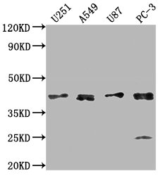
Western Blot Positive WB detected in: U251 whole cell lysate, A549 whole cell lysate, U87 whole cell lysate, PC-3 whole cell lysate All lanes: IKBIP antibody at 1:1000 Secondary Goat polyclonal to rabbit IgG at 1/50000 dilution Predicted band size: 40 kDa Observed band size: 40 kDa
Product group Antibodies
Cusabio
IKBIP AntibodyCSB-PA751229LA01HU
ApplicationsImmunoFluorescence, Western Blot, ELISA
ReactivityHuman
TargetIKBIP
  • SizePrice
Product group Antibodies
Cusabio
ApplicationsELISA
ReactivityHuman
TargetIKBIP
  • SizePrice