

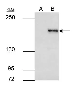
SETDB1 antibody [N2C1], Internal immunoprecipitates SETDB1 protein in IP experiments. IP samples: HeLa nuclear extract A. Control with 4 μg of preimmune Rabbit IgG B. Immunoprecipitation of SETDB1 protein by 4 μg SETDB1 antibody [N2C1], Internal (GTX110219) 5 % SDS-PAGE The immunoprecipitated SETDB1 protein was detected by SETDB1 antibody [N2C1], Internal (GTX110219) diluted at 1:1000. [EasyBlot anti-rabbit IgG (GTX221666-01) was used as a secondary reagent]
![SETDB1 antibody [N2C1], Internal detects SETDB1 protein at nucleus by immunofluorescent analysis. Sample: HeLa cells were fixed in 4% paraformaldehyde at RT for 15 min. Green: SETDB1 stained by SETDB1 antibody [N2C1], Internal (GTX110219) diluted at 1:500. Red: alpha Tubulin, a cytoskeleton marker, stained by alpha Tubulin antibody [GT114] (GTX628802) diluted at 1:1000. Scale bar= 10μm.](https://www.genetex.com/upload/website/prouct_img/normal/GTX110219/GTX110219_44504_20220408_ICC_IF_w_23060500_884.webp "SETDB1 antibody [N2C1], Internal detects SETDB1 protein at nucleus by immunofluorescent analysis. Sample: HeLa cells were fixed in 4% paraformaldehyde at RT for 15 min. Green: SETDB1 stained by SETDB1 antibody [N2C1], Internal (GTX110219) diluted at 1:500. Red: alpha Tubulin, a cytoskeleton marker, stained by alpha Tubulin antibody [GT114] (GTX628802) diluted at 1:1000. Scale bar= 10μm.")
![SETDB1 antibody [N2C1], Internal detects SETDB1 protein at nucleus by immunohistochemical analysis. Sample: Paraffin-embedded mouse testis. SETDB1 stained by SETDB1 antibody [N2C1], Internal (GTX110219) diluted at 1:500. Antigen Retrieval: Citrate buffer, pH 6.0, 15 min](https://www.genetex.com/upload/website/prouct_img/normal/GTX110219/GTX110219_44503_20220114_IHC-P_M_w_23060500_407.webp "SETDB1 antibody [N2C1], Internal detects SETDB1 protein at nucleus by immunohistochemical analysis. Sample: Paraffin-embedded mouse testis. SETDB1 stained by SETDB1 antibody [N2C1], Internal (GTX110219) diluted at 1:500. Antigen Retrieval: Citrate buffer, pH 6.0, 15 min")
![HeLa whole cell and nuclear extracts (30 μg) were separated by 5% SDS-PAGE, and the membrane was blotted with SETDB1 antibody [N2C1], Internal (GTX110219) diluted at 1:1000. The HRP-conjugated anti-rabbit IgG antibody (GTX213110-01) was used to detect the primary antibody.](https://www.genetex.com/upload/website/prouct_img/normal/GTX110219/GTX110219_40471_20210813_WB_Fraction_w_23060500_112.webp "HeLa whole cell and nuclear extracts (30 μg) were separated by 5% SDS-PAGE, and the membrane was blotted with SETDB1 antibody [N2C1], Internal (GTX110219) diluted at 1:1000. The HRP-conjugated anti-rabbit IgG antibody (GTX213110-01) was used to detect the primary antibody.")

SETDB1 antibody [N2C1], Internal immunoprecipitates SETDB1 protein in IP experiments. IP samples: HeLa nuclear extract A. Control with 4 μg of preimmune Rabbit IgG B. Immunoprecipitation of SETDB1 protein by 4 μg SETDB1 antibody [N2C1], Internal (GTX110219) 5 % SDS-PAGE The immunoprecipitated SETDB1 protein was detected by SETDB1 antibody [N2C1], Internal (GTX110219) diluted at 1:1000. [EasyBlot anti-rabbit IgG (GTX221666-01) was used as a secondary reagent]
SETDB1 antibody [N2C1], Internal
GTX110219
ApplicationsImmunoFluorescence, ImmunoPrecipitation, Western Blot, ChIP Chromatin ImmunoPrecipitation, ImmunoCytoChemistry, ImmunoHistoChemistry, ImmunoHistoChemistry Paraffin
Product group Antibodies
ReactivityHuman, Mouse
TargetSETDB1
Overview
- SupplierGeneTex
- Product NameSETDB1 antibody [N2C1], Internal
- Delivery Days Customer9
- Application Supplier NoteWB: 1:500-1:3000. IP: 1:100-1:500. *Optimal dilutions/concentrations should be determined by the researcher.Not tested in other applications.
- ApplicationsImmunoFluorescence, ImmunoPrecipitation, Western Blot, ChIP Chromatin ImmunoPrecipitation, ImmunoCytoChemistry, ImmunoHistoChemistry, ImmunoHistoChemistry Paraffin
- CertificationResearch Use Only
- ClonalityPolyclonal
- Concentration1 mg/ml
- ConjugateUnconjugated
- Gene ID9869
- Target nameSETDB1
- Target descriptionSET domain bifurcated histone lysine methyltransferase 1
- Target synonymsESET, H3-K9-HMTase4, KG1T, KMT1E, TDRD21, histone-lysine N-methyltransferase SETDB1, ERG-associated protein with a SET domain, ESET, SET domain bifurcated 1, histone H3-K9 methyltransferase 4, histone-lysine N-methyltransferase, H3lysine-9 specific 4, lysine N-methyltransferase 1E, tudor domain containing 21
- HostRabbit
- IsotypeIgG
- Protein IDQ15047
- Protein NameHistone-lysine N-methyltransferase SETDB1
- Scientific DescriptionThis gene encodes a histone methyltransferase. The encoded enzyme catalyzes the reaction of S-adenosyl-L-methionine and histone L-lysine to produce S-adenosyl-L-homocysteine and histone N(6)-methyl-L-lysine. The encoded protein likely functions in transcriptional repression. Alternatively spliced transcript variants have been described.
- ReactivityHuman, Mouse
- Storage Instruction-20°C or -80°C,2°C to 8°C
- UNSPSC41116161
Datasheet
Related products
Product group Antibodies
Anti-SETDB1 AntibodyA31125
ApplicationsWestern Blot, ImmunoHistoChemistry
ReactivityHuman, Mouse, Rat
- SizePrice
Product group Antibodies

Anti-SETDB1 [RAB-C531]AB01865-1.1-BT
ApplicationsImmunoFluorescence, ImmunoPrecipitation
ReactivityHuman
TargetSETDB1
- SizePrice
Product group Antibodies
Anti-SETDB1 Antibody144-06145
ApplicationsWestern Blot, ImmunoHistoChemistry
ReactivityHuman, Mouse, Rat
TargetSETDB1
- SizePrice
Product group Antibodies

ESET / SETDB1 AntibodyLS-C832291
ApplicationsELISA, ImmunoHistoChemistry
ReactivityHuman, Mouse
TargetSETDB1
- SizePrice
Product group Antibodies
SETDB1 Recombinant AntibodyBSM-60316M
ApplicationsWestern Blot
ReactivityHuman, Monkey, Mouse, Rat
TargetSETDB1
- SizePrice
Product group Antibodies
SETDB1 AntibodyCSB-PA619960ESR1HU
ApplicationsWestern Blot, ELISA, ImmunoHistoChemistry
ReactivityHuman, Rat
TargetSETDB1
- SizePrice
![ICC/IF analysis of LOVO cells using GTX80408 SETDB1 antibody [5H6A12]. Green : SETDB1 Red: Actin filaments](https://www.genetex.com/upload/website/prouct_img/normal/GTX80408/GTX80408_20170912_ICCIF_w_23061322_696.webp)
Product group Antibodies
SETDB1 antibody [5H6A12]GTX80408
ApplicationsImmunoFluorescence, Western Blot, ELISA, ImmunoCytoChemistry
ReactivityHuman, Monkey, Mouse
TargetSETDB1
- SizePrice
![WB analysis of MCF-7 (1),T47D (2), HEK293 (3), JURKAT (4), NIH3T3 (5) and F9 (6) cell lysate using GTX80409 SETDB1 antibody [5H6D4].](https://www.genetex.com/upload/website/prouct_img/normal/GTX80409/GTX80409_20170912_WB_w_23061322_503.webp)
Product group Antibodies
SETDB1 antibody [5H6D4]GTX80409
ApplicationsWestern Blot, ELISA
ReactivityHuman, Monkey, Mouse
TargetSETDB1
- SizePrice
![Whole cell extract (30 μg) was separated by 5% SDS-PAGE, and the membrane was blotted with SETDB1 antibody [N1], N-term (GTX115305) diluted at 1:2000. The HRP-conjugated anti-rabbit IgG antibody (GTX213110-01) was used to detect the primary antibody.](https://www.genetex.com/upload/website/prouct_img/normal/GTX115305/GTX115305_40604_20230224_WB_23030219_547.webp)
Product group Antibodies
SETDB1 antibody [N1], N-termGTX115305
ApplicationsImmunoPrecipitation, Western Blot, ChIP Chromatin ImmunoPrecipitation, ImmunoHistoChemistry, ImmunoHistoChemistry Paraffin
ReactivityHuman, Mouse
TargetSETDB1
- SizePrice