

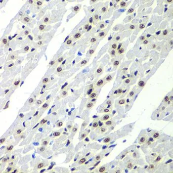
IHC-P analysis of rat heart tissue using GTX32869 SF3B2 antibody. Dilution : 1:100


IHC-P analysis of rat heart tissue using GTX32869 SF3B2 antibody. Dilution : 1:100
SF3B2 antibody
GTX32869
ApplicationsImmunoFluorescence, Western Blot, ImmunoCytoChemistry, ImmunoHistoChemistry, ImmunoHistoChemistry Paraffin
Product group Antibodies
ReactivityHuman, Mouse, Rat
TargetSF3B2
Overview
- SupplierGeneTex
- Product NameSF3B2 antibody
- Delivery Days Customer9
- Application Supplier NoteWB: 1:500 - 1:2000. ICC/IF: 1:50 - 1:200. IHC-P: 1:50 - 1:200. *Optimal dilutions/concentrations should be determined by the researcher.Not tested in other applications.
- ApplicationsImmunoFluorescence, Western Blot, ImmunoCytoChemistry, ImmunoHistoChemistry, ImmunoHistoChemistry Paraffin
- CertificationResearch Use Only
- ClonalityPolyclonal
- ConjugateUnconjugated
- Gene ID10992
- Target nameSF3B2
- Target descriptionsplicing factor 3b subunit 2
- Target synonymsCFM, Cus1, HFM, SAP145, SF3B145, SF3b1, SF3b150, splicing factor 3B subunit 2, SAP 145, pre-mRNA splicing factor SF3b 145 kDa subunit, spliceosome associated protein 145, splicing factor 3b, subunit 2, 145kD, splicing factor 3b, subunit 2, 145kDa
- HostRabbit
- IsotypeIgG
- Protein IDQ13435
- Protein NameSplicing factor 3B subunit 2
- Scientific DescriptionThis gene encodes subunit 2 of the splicing factor 3b protein complex. Splicing factor 3b, together with splicing factor 3a and a 12S RNA unit, forms the U2 small nuclear ribonucleoproteins complex (U2 snRNP). The splicing factor 3b/3a complex binds pre-mRNA upstream of the introns branch site in a sequence-independent manner and may anchor the U2 snRNP to the pre-mRNA. Splicing factor 3b is also a component of the minor U12-type spliceosome. Subunit 2 associates with pre-mRNA upstream of the branch site at the anchoring site. Subunit 2 also interacts directly with subunit 4 of the splicing factor 3b complex. Subunit 2 is a highly hydrophilic protein with a proline-rich N-terminus and a glutamate-rich stretch in the C-terminus. [provided by RefSeq, Jul 2008]
- ReactivityHuman, Mouse, Rat
- Storage Instruction-20°C or -80°C,2°C to 8°C
- UNSPSC41116161
Datasheet
Related products
Product group Antibodies
SF3B2 AntibodyCSB-PA615687DSR2HU
ApplicationsELISA, ImmunoHistoChemistry
ReactivityHuman
TargetSF3B2
- SizePrice

Product group Antibodies
Anti-SF3B2 Antibody Picoband(r)A07047-1-CARRIER-FREE
ApplicationsFlow Cytometry, Western Blot, ELISA
ReactivityHuman, Mouse, Rat
TargetSF3B2
- SizePrice
Product group Antibodies
Anti-SF3B2 AntibodyA31236
ApplicationsImmunoFluorescence, Western Blot, ImmunoHistoChemistry
ReactivityHuman, Mouse, Rat
- SizePrice
Product group Antibodies
Anti-SF3B2 AntibodyHPA045028
ApplicationsWestern Blot, ImmunoCytoChemistry, ImmunoHistoChemistry
ReactivityHuman
TargetSF3B2
- SizePrice
Product group Antibodies

SF3B2 AntibodyLS-C334356
ApplicationsImmunoFluorescence, Western Blot, ImmunoHistoChemistry
ReactivityHuman, Mouse, Rat
TargetSF3B2
- SizePrice
Product group Antibodies
Anti-SF3B2 Antibody144-05875
ApplicationsImmunoFluorescence, Western Blot, ImmunoHistoChemistry
ReactivityHuman, Mouse, Rat
TargetSF3B2
- SizePrice