

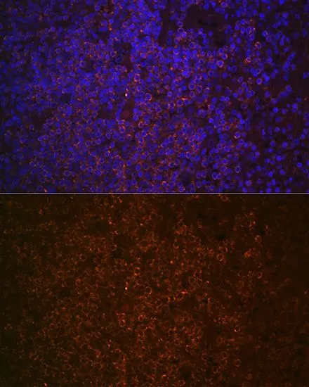
IHC-P analysis of human spleen tissue section using GTX03199 SHIP1 antibody [GT1287]. Blue : DAPI for nuclear staining Dilution : 1:100
![WB analysis of various samples using GTX03199 SHIP1 antibody [GT1287]. Dilution : 1:1000 Loading : 25μg per lane](https://www.genetex.com/upload/website/prouct_img/normal/GTX03199/GTX03199_23_WB_w_23053123_802.webp "WB analysis of various samples using GTX03199 SHIP1 antibody [GT1287]. Dilution : 1:1000 Loading : 25μg per lane")
![IHC-P analysis of rat spleen tissue section using GTX03199 SHIP1 antibody [GT1287]. Blue : DAPI for nuclear staining Dilution : 1:100](https://www.genetex.com/upload/website/prouct_img/normal/GTX03199/GTX03199_20210615_IHC-P_19_w_23053123_492.webp "IHC-P analysis of rat spleen tissue section using GTX03199 SHIP1 antibody [GT1287]. Blue : DAPI for nuclear staining Dilution : 1:100")
![IHC-P analysis of human spleen tissue section using GTX03199 SHIP1 antibody [GT1287]. Dilution : 1:100](https://www.genetex.com/upload/website/prouct_img/normal/GTX03199/GTX03199_20210615_IHC-P_18_w_23053123_352.webp "IHC-P analysis of human spleen tissue section using GTX03199 SHIP1 antibody [GT1287]. Dilution : 1:100")
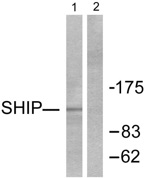
![Various whole cell extracts (30 μg) were separated by 5% SDS-PAGE, and the membrane was blotted with SHIP1 antibody [GT1287] (GTX03199) diluted at 1:1000. The HRP-conjugated anti-rabbit IgG antibody (GTX213110-01) was used to detect the primary antibody.](https://www.genetex.com/upload/website/prouct_img/normal/GTX03199/GTX03199_4000002047_20210709_WB_w_23053123_792.webp "Various whole cell extracts (30 μg) were separated by 5% SDS-PAGE, and the membrane was blotted with SHIP1 antibody [GT1287] (GTX03199) diluted at 1:1000. The HRP-conjugated anti-rabbit IgG antibody (GTX213110-01) was used to detect the primary antibody.")
![IHC-P analysis of mouse spleen tissue section using GTX03199 SHIP1 antibody [GT1287]. Blue : DAPI for nuclear staining Dilution : 1:100](https://www.genetex.com/upload/website/prouct_img/normal/GTX03199/GTX03199_20210615_IHC-P_21_w_23053123_970.webp "IHC-P analysis of mouse spleen tissue section using GTX03199 SHIP1 antibody [GT1287]. Blue : DAPI for nuclear staining Dilution : 1:100")
![Various whole cell extracts (30 μg) were separated by 5% SDS-PAGE, and the membrane was blotted with SHIP1 antibody [GT1287] (GTX03199) diluted at 1:1000. The HRP-conjugated anti-rabbit IgG antibody (GTX213110-01) was used to detect the primary antibody.](https://www.genetex.com/upload/website/prouct_img/normal/GTX03199/GTX03199_4000002047_20210709_WB_2_w_23053123_513.webp "Various whole cell extracts (30 μg) were separated by 5% SDS-PAGE, and the membrane was blotted with SHIP1 antibody [GT1287] (GTX03199) diluted at 1:1000. The HRP-conjugated anti-rabbit IgG antibody (GTX213110-01) was used to detect the primary antibody.")
IHC-P analysis of human spleen tissue section using GTX03199 SHIP1 antibody [GT1287]. Blue : DAPI for nuclear staining Dilution : 1:100
SHIP1 antibody [GT1287]
GTX03199
ApplicationsImmunoFluorescence, ImmunoPrecipitation, Western Blot, ImmunoCytoChemistry, ImmunoHistoChemistry, ImmunoHistoChemistry Paraffin
Product group Antibodies
ReactivityHuman, Mouse, Rat
TargetINPP5D
Overview
- SupplierGeneTex
- Product NameSHIP1 antibody [GT1287]
- Delivery Days Customer9
- Application Supplier NoteWB: 1:500 - 1:2000. ICC/IF: 1:50 - 1:200. IHC-P: 1:50 - 1:200. *Optimal dilutions/concentrations should be determined by the researcher.Not tested in other applications.
- ApplicationsImmunoFluorescence, ImmunoPrecipitation, Western Blot, ImmunoCytoChemistry, ImmunoHistoChemistry, ImmunoHistoChemistry Paraffin
- CertificationResearch Use Only
- ClonalityMonoclonal
- Clone IDGT1287
- Concentration0.79 mg/ml
- ConjugateUnconjugated
- Gene ID3635
- Target nameINPP5D
- Target descriptioninositol polyphosphate-5-phosphatase D
- Target synonymsSHIP, SHIP-1, SHIP1, SIP-145, hp51CN, p150Ship, phosphatidylinositol 3,4,5-trisphosphate 5-phosphatase 1, SH2 domain-containing inositol 5'-phosphatase 1, inositol polyphosphate-5-phosphatase, 145kD, inositol polyphosphate-5-phosphatase, 145kDa, phosphatidylinositol 4,5-bisphosphate 5-phosphatase, signaling inositol polyphosphate 5 phosphatase SIP-145, signaling inositol polyphosphate phosphatase SHIP II
- HostRabbit
- IsotypeIgG
- Protein IDQ92835
- Protein NamePhosphatidylinositol 3,4,5-trisphosphate 5-phosphatase 1
- Scientific DescriptionThis gene is a member of the inositol polyphosphate-5-phosphatase (INPP5) family and encodes a protein with an N-terminal SH2 domain, an inositol phosphatase domain, and two C-terminal protein interaction domains. Expression of this protein is restricted to hematopoietic cells where its movement from the cytosol to the plasma membrane is mediated by tyrosine phosphorylation. At the plasma membrane, the protein hydrolyzes the 5 phosphate from phosphatidylinositol (3,4,5)-trisphosphate and inositol-1,3,4,5-tetrakisphosphate, thereby affecting multiple signaling pathways. The protein is also partly localized to the nucleus, where it may be involved in nuclear inositol phosphate signaling processes. Overall, the protein functions as a negative regulator of myeloid cell proliferation and survival. Mutations in this gene are associated with defects and cancers of the immune system. Alternative splicing of this gene results in multiple transcript variants. [provided by RefSeq, Feb 2014]
- ReactivityHuman, Mouse, Rat
- Storage Instruction-20°C or -80°C,2°C to 8°C
- UNSPSC41116161
Datasheet
Related products
Product group Antibodies
Anti-SHIP1 AntibodyA97231
ApplicationsWestern Blot, ELISA
ReactivityHuman, Mouse, Rat
- SizePrice

Product group Antibodies
Anti-INPP5D Antibody Picoband(r)A03358-1-CARRIER-FREE
ApplicationsFlow Cytometry, Western Blot, ELISA
ReactivityHuman
TargetINPP5D
- SizePrice
Product group Antibodies
Anti-Phospho-INPP5D-Y1020 Antibody144-50772
ApplicationsWestern Blot
ReactivityHuman
TargetINPP5D
- SizePrice
Product group Antibodies
References
ApplicationsFlow Cytometry, Western Blot, ELISA
ReactivityBovine, Canine, Human, Mouse, Porcine, Rat
TargetINPP5D
- SizePrice
Product group Antibodies
INPP5D AntibodyCSB-PA314107
ApplicationsWestern Blot, ELISA
ReactivityHuman, Mouse, Rat
TargetINPP5D
- SizePrice
Product group Antibodies
Inpp5D Recombinant AntibodyCAC12573
ApplicationsImmunoPrecipitation, Western Blot, ELISA, ImmunoHistoChemistry
TargetINPP5D
- SizePrice
Product group Antibodies

ApplicationsWestern Blot
ReactivityBovine, Human, Mouse, Rat
TargetINPP5D
- SizePrice
![WB analysis of THP-1 cells using GTX79893 SHIP1 antibody [SHIP-02]. Lane 1 : mouse IgG1 isotype control Lane 2,3 : GTX79894 Lane 4,5 : GTX79893](https://www.genetex.com/upload/website/prouct_img/normal/GTX79893/GTX79893_20191025_AP_001_322_w_23061322_633.webp)
Product group Antibodies
SHIP1 antibody [SHIP-02]GTX79893
ApplicationsFlow Cytometry, ImmunoFluorescence, Western Blot, ImmunoCytoChemistry
ReactivityHuman
TargetINPP5D
- SizePrice
![WB analysis of THP-1 cells using GTX79894 SHIP1 antibody [SHIP-01]. Lane 1 : mouse IgG1 isotype control Lane 2,3 : GTX79894 Lane 4,5 : GTX79893](https://www.genetex.com/upload/website/prouct_img/normal/GTX79894/GTX79894_20191025_AP_001_323_w_23061322_181.webp)
Product group Antibodies
SHIP1 antibody [SHIP-01]GTX79894
ApplicationsFlow Cytometry, Western Blot
ReactivityHuman
TargetINPP5D
- SizePrice
Product group Antibodies
Anti-INPP5D AntibodyHPA070455
ApplicationsWestern Blot, ImmunoCytoChemistry, ImmunoHistoChemistry
ReactivityHuman
TargetINPP5D
- SizePrice