

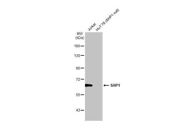
Various whole cell extracts (30 μg) were separated by 7.5% SDS-PAGE, and the membrane was blotted with SHP1 antibody [HL20845] (GTX637993) diluted at 1:1000. The HRP-conjugated anti-rabbit IgG antibody (GTX213110-01) was used to detect the primary antibody.
![Various whole cell extracts (30 μg) were separated by 7.5% SDS-PAGE, and the membrane was blotted with SHP1 antibody [HL20845] (GTX637993) diluted at 1:1000. The HRP-conjugated anti-rabbit IgG antibody (GTX213110-01) was used to detect the primary antibody. Corresponding RNA expression data for the same cell lines are based on Human Protein Atlas program.](https://www.genetex.com/upload/website/prouct_img/normal/GTX637993/GTX637993_44970_20230303_WB_TPM_watermark_23030717_704.webp "Various whole cell extracts (30 μg) were separated by 7.5% SDS-PAGE, and the membrane was blotted with SHP1 antibody [HL20845] (GTX637993) diluted at 1:1000. The HRP-conjugated anti-rabbit IgG antibody (GTX213110-01) was used to detect the primary antibody. Corresponding RNA expression data for the same cell lines are based on Human Protein Atlas program.")
![SHP1 antibody [HL20845] detects SHP1 protein at cytoplasm by immunofluorescent analysis. Sample: Jurkat cells were fixed in 4% paraformaldehyde at RT for 15 min. Green: SHP1 stained by SHP1 antibody [HL20845] (GTX637993) diluted at 1:500. Blue: Fluoroshield with DAPI (GTX30920).](https://www.genetex.com/upload/website/prouct_img/normal/GTX637993/GTX637993_44970_20230428_ICC_IF_23050918_365.webp "SHP1 antibody [HL20845] detects SHP1 protein at cytoplasm by immunofluorescent analysis. Sample: Jurkat cells were fixed in 4% paraformaldehyde at RT for 15 min. Green: SHP1 stained by SHP1 antibody [HL20845] (GTX637993) diluted at 1:500. Blue: Fluoroshield with DAPI (GTX30920).")
![SHP1 antibody [HL20845] detects SHP1 protein at cytoplasm by immunofluorescent analysis. Sample: MCF-7 cells were fixed in 4% paraformaldehyde at RT for 15 min. Green: SHP1 stained by SHP1 antibody [HL20845] (GTX637993) diluted at 1:500. Blue: Fluoroshield with DAPI (GTX30920).](https://www.genetex.com/upload/website/prouct_img/normal/GTX637993/GTX637993_44970_20230526_ICC_IF_23060622_517.webp "SHP1 antibody [HL20845] detects SHP1 protein at cytoplasm by immunofluorescent analysis. Sample: MCF-7 cells were fixed in 4% paraformaldehyde at RT for 15 min. Green: SHP1 stained by SHP1 antibody [HL20845] (GTX637993) diluted at 1:500. Blue: Fluoroshield with DAPI (GTX30920).")
![Whole cell extract (30 μg) was separated by 7.5% SDS-PAGE, and the membranes were blotted with SHP1 antibody [HL20845] (GTX637993) diluted at 1:1000 and competitor's antibody (#Highly competitor antibody) diluted at 1:1000. The HRP-conjugated anti-rabbit IgG antibody (GTX213110-01) was used to detect the primary antibody. *The competitor is not affiliated with GeneTex and does not endorse this product.](https://www.genetex.com/upload/website/prouct_img/normal/GTX637993/GTX637993_44970_20230707_WB_competitor_watermark_23071223_813.webp "Whole cell extract (30 μg) was separated by 7.5% SDS-PAGE, and the membranes were blotted with SHP1 antibody [HL20845] (GTX637993) diluted at 1:1000 and competitor's antibody (#Highly competitor antibody) diluted at 1:1000. The HRP-conjugated anti-rabbit IgG antibody (GTX213110-01) was used to detect the primary antibody. *The competitor is not affiliated with GeneTex and does not endorse this product.")
Various whole cell extracts (30 μg) were separated by 7.5% SDS-PAGE, and the membrane was blotted with SHP1 antibody [HL20845] (GTX637993) diluted at 1:1000. The HRP-conjugated anti-rabbit IgG antibody (GTX213110-01) was used to detect the primary antibody.
SHP1 antibody [HL20845]
GTX637993
ApplicationsImmunoFluorescence, Western Blot, ImmunoCytoChemistry
Product group Antibodies
ReactivityHuman
TargetPTPN6
Overview
- SupplierGeneTex
- Product NameSHP1 antibody [HL20845]
- Delivery Days Customer9
- Application Supplier NoteWB: 1:500-1:3000. *Optimal dilutions/concentrations should be determined by the researcher.Not tested in other applications.
- ApplicationsImmunoFluorescence, Western Blot, ImmunoCytoChemistry
- CertificationResearch Use Only
- ClonalityMonoclonal
- Clone IDHL20845
- Concentration1 mg/ml
- ConjugateUnconjugated
- Gene ID5777
- Target namePTPN6
- Target descriptionprotein tyrosine phosphatase non-receptor type 6
- Target synonymsHCP, HCPH, HPTP1C, PTP-1C, SH-PTP1, SHP-1, SHP-1L, SHP1, tyrosine-protein phosphatase non-receptor type 6, hematopoietic cell phosphatase, hematopoietic cell protein-tyrosine phosphatase, protein-tyrosine phosphatase 1C, protein-tyrosine phosphatase SHP-1
- HostRabbit
- IsotypeIgG
- Protein IDP29350
- Protein NameTyrosine-protein phosphatase non-receptor type 6
- Scientific DescriptionThe protein encoded by this gene is a member of the protein tyrosine phosphatase (PTP) family. PTPs are known to be signaling molecules that regulate a variety of cellular processes including cell growth, differentiation, mitotic cycle, and oncogenic transformation. N-terminal part of this PTP contains two tandem Src homolog (SH2) domains, which act as protein phospho-tyrosine binding domains, and mediate the interaction of this PTP with its substrates. This PTP is expressed primarily in hematopoietic cells, and functions as an important regulator of multiple signaling pathways in hematopoietic cells. This PTP has been shown to interact with, and dephosphorylate a wide spectrum of phospho-proteins involved in hematopoietic cell signaling. Multiple alternatively spliced variants of this gene, which encode distinct isoforms, have been reported. [provided by RefSeq, Jul 2008]
- ReactivityHuman
- Storage Instruction-20°C or -80°C,2°C to 8°C
- UNSPSC41116161
Datasheet
Related products
Product group Antibodies
PTPN6 AntibodyCSB-PA004085
ApplicationsWestern Blot, ELISA
ReactivityHuman
TargetPTPN6
- SizePrice
Product group Antibodies
PTPN6 Polyclonal AntibodyCAC13888
ApplicationsImmunoFluorescence, Western Blot, ELISA, ImmunoHistoChemistry
TargetPTPN6
- SizePrice
Product group Antibodies
Anti-SHP-1 AntibodyA100768
ApplicationsWestern Blot, ELISA
ReactivityHuman
- SizePrice
Product group Antibodies
Anti-PTPN6 Antibody144-01446
ApplicationsImmunoFluorescence, Western Blot
ReactivityHuman, Mouse
TargetPTPN6
- SizePrice

Product group Antibodies
Anti-SHP1/PTPN6 Antibody Picoband(r)A00938-3-CARRIER-FREE
ApplicationsFlow Cytometry, ImmunoFluorescence, Western Blot, ELISA, ImmunoCytoChemistry
ReactivityHuman, Mouse
TargetPTPN6
- SizePrice
Product group Antibodies

ApplicationsWestern Blot, ELISA
ReactivityHuman
TargetPTPN6
- SizePrice

Product group Antibodies
Anti-PTPN6 AntibodyHPA001466
ApplicationsWestern Blot, ImmunoCytoChemistry, ImmunoHistoChemistry
ReactivityHuman, Rat
TargetPTPN6
- SizePrice
Product group Antibodies

PTPN6 / SHP1 AntibodyLS-C401020
ApplicationsWestern Blot, ELISA, ImmunoHistoChemistry
ReactivityHuman, Mouse, Rat
TargetPTPN6
- SizePrice
Product group Antibodies
SHP1 (phospho Tyr536) antibodyGTX78946
ApplicationsImmunoHistoChemistry, ImmunoHistoChemistry Paraffin
ReactivityHuman
TargetPTPN6
- SizePrice