

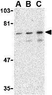
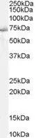
WB analysis of mouse skeletal muscle tissue lysate using GTX31744 SHP2 antibody. Working concentration : (A) 0.5, (B) 1, and (C) 2 μg/ml
WB analysis of mouse skeletal muscle tissue lysate using GTX31744 SHP2 antibody. Working concentration : (A) 0.5, (B) 1, and (C) 2 μg/ml
SHP2 antibody
GTX31744
ApplicationsWestern Blot, ELISA
Product group Antibodies
ReactivityHuman, Mouse, Rat
TargetPTPN11
Overview
- SupplierGeneTex
- Product NameSHP2 antibody
- Delivery Days Customer9
- Application Supplier NoteWB: 0.5 - 2 microg/mL. *Optimal dilutions/concentrations should be determined by the researcher.Not tested in other applications.
- ApplicationsWestern Blot, ELISA
- CertificationResearch Use Only
- ClonalityPolyclonal
- Concentration1 mg/ml
- ConjugateUnconjugated
- Gene ID5781
- Target namePTPN11
- Target descriptionprotein tyrosine phosphatase non-receptor type 11
- Target synonymsBPTP3, CFC, JMML, METCDS, NS1, PTP-1D, PTP2C, SH-PTP2, SH-PTP3, SHP2, tyrosine-protein phosphatase non-receptor type 11, SH2 domain-containing protein tyrosine phosphatase 2, protein-tyrosine phosphatase 1D, protein-tyrosine phosphatase 2C
- HostRabbit
- IsotypeIgG
- Protein IDQ06124
- Protein NameTyrosine-protein phosphatase non-receptor type 11
- Scientific DescriptionThe protein encoded by this gene is a member of the protein tyrosine phosphatase (PTP) family. PTPs are known to be signaling molecules that regulate a variety of cellular processes including cell growth, differentiation, mitotic cycle, and oncogenic transformation. This PTP contains two tandem Src homology-2 domains, which function as phospho-tyrosine binding domains and mediate the interaction of this PTP with its substrates. This PTP is widely expressed in most tissues and plays a regulatory role in various cell signaling events that are important for a diversity of cell functions, such as mitogenic activation, metabolic control, transcription regulation, and cell migration. Mutations in this gene are a cause of Noonan syndrome as well as acute myeloid leukemia. Two transcript variants encoding different isoforms have been found for this gene. [provided by RefSeq, May 2012]
- ReactivityHuman, Mouse, Rat
- Storage Instruction-20°C or -80°C,2°C to 8°C
- UNSPSC41116161
Datasheet
Related products
Product group Antibodies
Anti-PTPN11 Antibody144-65590
ApplicationsImmunoFluorescence, Western Blot, ImmunoHistoChemistry
ReactivityHuman, Mouse, Rat
TargetPTPN11
- SizePrice
Product group Antibodies
ApplicationsFlow Cytometry, Western Blot, ImmunoCytoChemistry
ReactivityHuman, Mouse, Rat
TargetPTPN11
- SizePrice
Product group Antibodies
PTPN11 AntibodyCSB-PA004086
ApplicationsWestern Blot, ELISA, ImmunoHistoChemistry
ReactivityHuman, Mouse, Rat
TargetPTPN11
- SizePrice
Product group Antibodies

Goat anti-SHP2 / PTPN11EB05114
ApplicationsFlow Cytometry, ImmunoFluorescence, Western Blot, ELISA
ReactivityBovine, Human, Mouse, Porcine, Rat
TargetPTPN11
- SizePrice
Product group Antibodies
ApplicationsImmunoPrecipitation, Western Blot, ELISA
TargetPTPN11
- SizePrice
Product group Antibodies

PTPN11 / SHP-2 / NS1 AntibodyLS-C404276
ApplicationsWestern Blot, ELISA, ImmunoHistoChemistry
ReactivityHuman, Mouse, Rat
TargetPTPN11
- SizePrice
Product group Antibodies
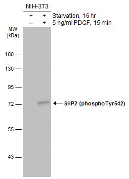
SHP2 (phospho Tyr542) antibodyGTX133983
ApplicationsWestern Blot
ReactivityHuman, Mouse
TargetPTPN11
- SizePrice
Product group Antibodies
SHP2 (phospho Tyr542) antibodyGTX17939
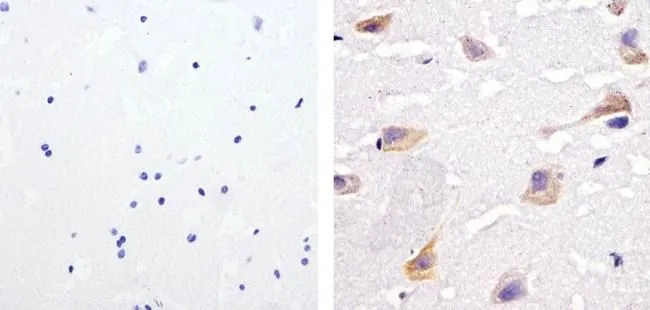
ApplicationsWestern Blot, ImmunoHistoChemistry, ImmunoHistoChemistry Paraffin
ReactivityHuman, Mouse
TargetPTPN11
- SizePrice
Product group Antibodies
References
SHP2 antibody, C-termGTX29214
ApplicationsWestern Blot, ImmunoHistoChemistry, ImmunoHistoChemistry Paraffin
ReactivityHuman, Mouse
TargetPTPN11
- SizePrice